Transcarotid Transcatheter Aortic Valve Replacement as Preferred Alternative Access in a Patient With Bilateral Carotid Artery Disease
J INVASIVE CARDIOL 2018;30(1):E9-E10.
Key words: aortic stenosis, TAVR, transcarotid access
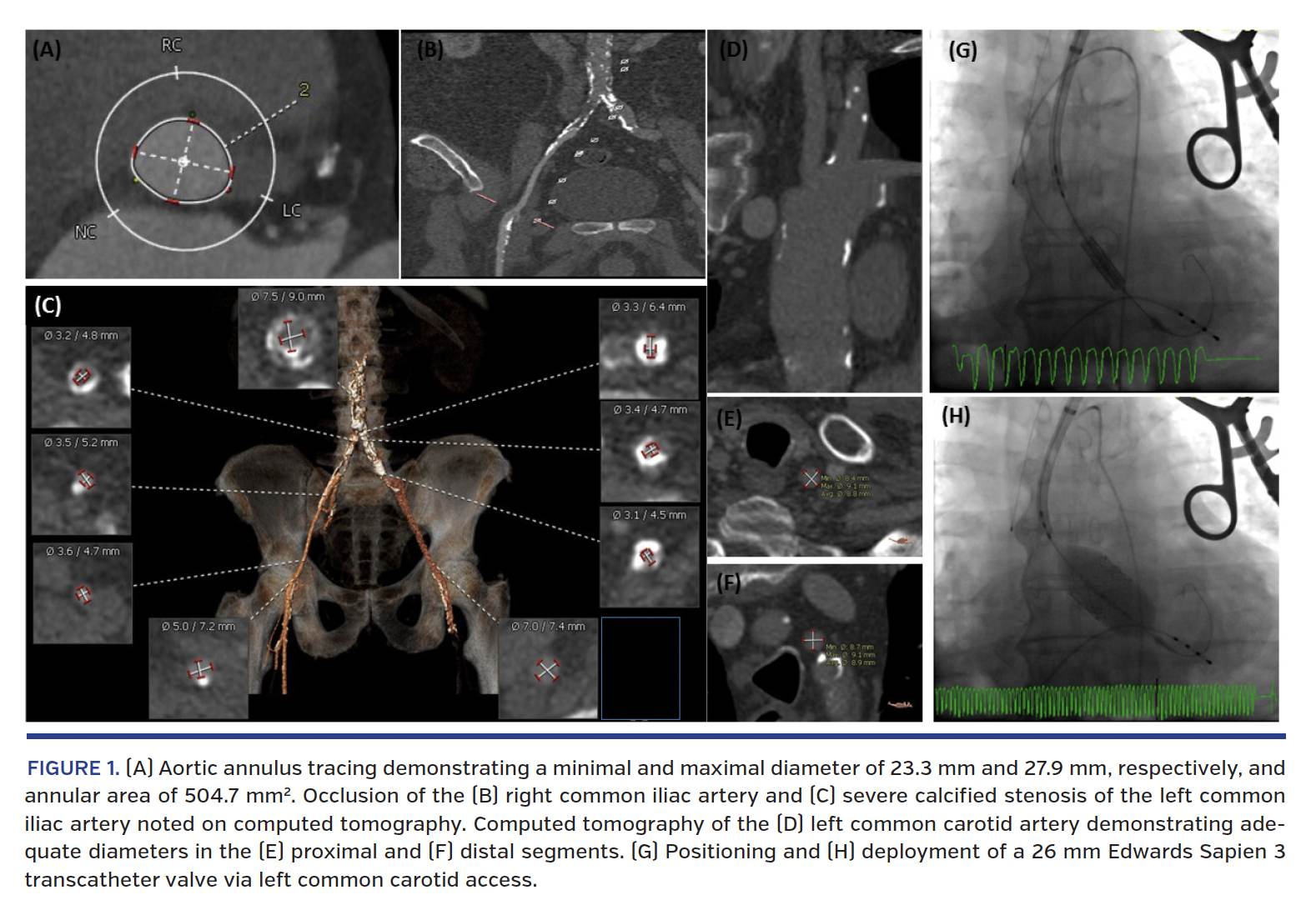
A frail 78-year-old man presented with severe symptomatic aortic stenosis. Echocardiography revealed a heavily calcified and stenotic aortic valve (Figure 1A), with an area of 0.75 cm2 and a mean gradient of 43 mm Hg. Given multiple comorbidities and his frailty status, the heart team concurred that a transcatheter approach was most suitable. Computed tomography angiography revealed poor transfemoral access due to an occluded right common iliac artery (Figure 1B) and a severely calcified left common iliac artery (Figure 1C) with minimal luminal diameter of 3.1 mm. Carotid ultrasound demonstrated an occluded left internal carotid artery (ICA) and a severely stenotic right ICA. Given the left ICA was completely occluded, a decision was made to pursue transcatheter aortic valve replacement (TAVR) via left common carotid artery (CCA) access (Figures 1D-1F).
Left CCA cutdown was performed and a 10 mm Hemashield graft (Maquet) was attached. Left femoral artery and vein were accessed for insertion of a pigtail catheter and a transvenous pacer, respectively. A 7 Fr sheath was inserted into the Hemashield graft. A diagnostic catheter and wire were inserted into the left ventricle. A stiff wire was placed into the left ventricle. A 14 Fr Edwards sheath (Edwards Lifesciences) was then advanced over the stiff wire such that the distal tip of the sheath was just below the takeoff of the left CCA in the arch (Figure 1G). A 26 mm Sapien 3 valve (Edwards Lifesciences) was then introduced (Figure 1H). The nosecone of the delivery system was advanced to the aortic annulus, and docking and alignment of the valve markers were performed in the ascending aorta. The valve was advanced to the aortic annulus and angiograms were performed to adjust position. The valve was successfully deployed with rapid pacing. Echocardiogram demonstrated optimal valve position with no paravalvular leak. The delivery system and wire were removed from the sheath. The sheath was then removed and the graft was stapled off. The incision was closed. The patient was extubated in the catheterization laboratory. A blood pressure of 140-150 mm Hg was maintained throughout (except at the time of rapid pacing) to ensure adequate cerebral perfusion pressure. The patient was discharged home 4 days later with symptomatic improvement.
While transcarotid TAVR has previously been described,1-3 to our knowledge, it has not been reported in patients with bilateral carotid disease. This case illustrates that in patients with an occluded ICA and a severely stenotic contralateral ICA, ipsilateral transcarotid access is safe and feasible and may be the preferred alternative access for TAVR.

References
1. Kirker EB, Hodson RW, Spinelli KJ, Korngold EC. The carotid artery as a preferred alternative access route for transcatheter aortic valve replacement. Ann Thorac Surg. 2017;104:621-629. Epub 2017 Mar 6.
2. Mylotte D, Sudre A, Teiger E, et al. Transcarotid transcatheter aortic valve replacement: feasibility and safety. JACC Cardiovasc Interv. 2016;9:472-480.
3. Guyton RA, Block PC, Thourani VH, Lerakis S, Babaliaros V. Carotid artery access for transcatheter aortic valve replacement. Catheter Cardiovasc Interv. 2013;82:E583-E586.
From the 1Department of Medicine and 2Department of Surgery, Stony Brook University Hospital, Stony Brook, New York.
Disclosure: The authors have completed and returned the ICMJE Form for Disclosure of Potential Conflicts of Interest. The authors report no conflicts of interest regarding the content herein.
Manuscript accepted April 9, 2017.
Address for correspondence: Puja B. Parikh, MD, MPH, FACC, FAHA, FSCAI, Co-Director, Transcatheter Aortic Valve Replacement Program, Stony Brook University Medical Center, Health Sciences Center T16-080, Stony Brook, NY 11794-8160. Email: puja.parikh@stonybrookmedicine.edu


