Spontaneously Sealed Forearm Radial Artery Perforation During a Left Distal Transradial Coronary Intervention
Key words: cardiac imaging, distal transradial intervention, radial artery perforation
The adoption of distal transradial access (dTRA) as the default approach for coronary angiography and interventions was recently published. As a refinement of conventional (proximal) TRA, this technique has advantages in terms of patient and operator comfort and risk of radial artery occlusion. Radial artery perforation (RAP) is a rare but dreaded complication of any TRA, which should therefore be promptly recognized and managed in order to avoid compartment syndrome.
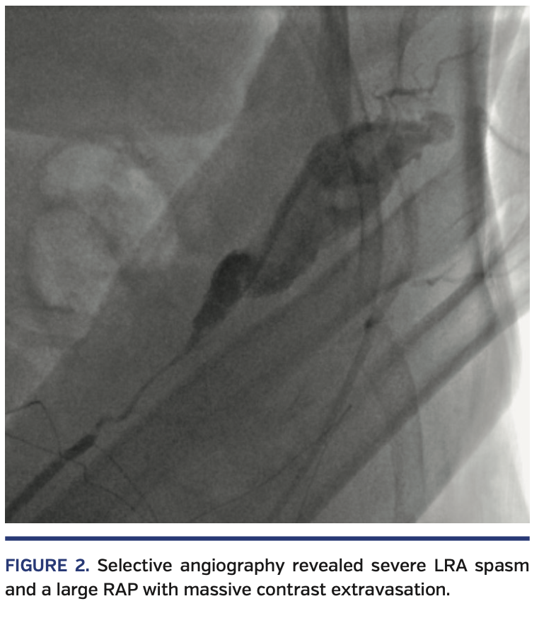
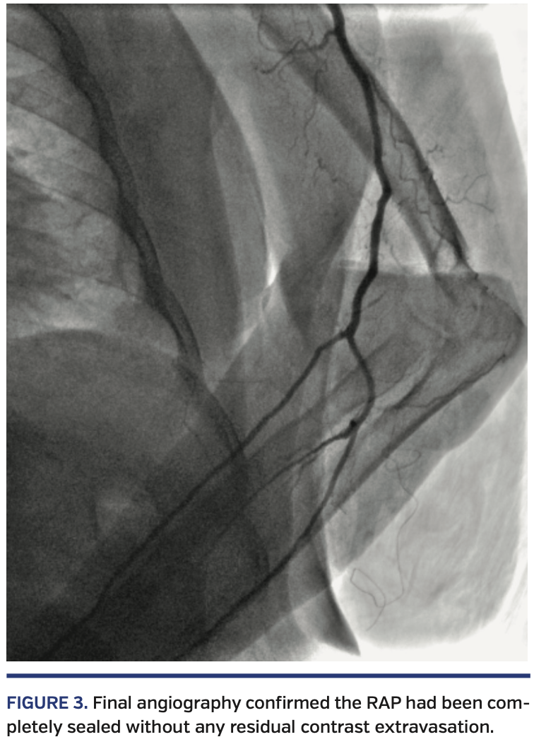
A 62-year-old woman with short stature, hypertension, diabetes, obesity, peripheral arterial disease, and previous acute myocardial infarction (AMI) requiring percutaneous coronary intervention (PCI) of the left anterior descending artery presented with non-ST elevation AMI. She was referred to urgent coronary angiography via left dTRA, using a 5/6 Fr hydrophilic slender Glidesheath (Terumo) inserted without any difficulties (Figure 1). Despite intra-arterial nitroglycerin, resistance was felt at the level of the forearm left radial artery (LRA), and it was not possible to further advance even a hydrophilic 0.035˝ J-tip wire through the LRA. Selective angiography (through the sheath) revealed severe LRA spasm and a large RAP with massive contrast extravasation (Figure 2; Video 1). The wire-crossing attempts always resulted in the false lumen; it was therefore decided to cross the site of perforation with a 5 Fr MP diagnostic catheter, without the rail of a wire, by gentle rotations and advancements. By confirming the normal arterial waveform pressure just proximal to the perforated area, the system was then carefully advanced up to the aortic root. The dominant right coronary artery (RCA) was diffusely diseased, with proximal-mid critical stenosis and a consequent Thrombolysis in Myocardial Infarction 2 flow (Video 2), without any significant lesions at the left coronary. The culprit RCA was promptly fixed by PCI with 3 drug-eluting stents (distal to ostial), requiring adequate pre- and postdilations and laborious reversion of an adenosine refractory no-reflow with papaverine (Video 3). By withdrawing the JR4 6 Fr guiding catheter, a final angiogram confirmed the perforation had been completely sealed without any residual contrast extravasation (Figure 3; Video 4). The patient was monitored for any signs of forearm compromise and uneventfully discharged home 2 days later.
RAP is an unusual but threatening complication that can be conservatively managed with guide-catheter tamponade by performing the intended procedure through the same TRA.
View the Accompanying Video Series Here.
From the Department of Interventional Cardiology, Hospital São Paulo, Escola Paulista de Medicina, Universidade Federal de São Paulo, São Paulo, Brazil.
Disclosure: The authors have completed and returned the ICMJE Form for Disclosure of Potential Conflicts of Interest. The authors report no conflicts of interest regarding the content herein.
The authors report that patient consent was provided for publication of the images used herein.
Manuscript accepted February 5, 2020.
Address for correspondence: Marcos Danillo Peixoto Oliveira, MD, Napoleão de Barros street, nº 715 - Vila Clementino, Sao Paulo-SP, Brazil, 04024-002. Email: mdmarcosdanillo@gmail.com


