Endovascular Treatment of a Migrated Superior Vena Cava Stent in the Right Atrium
J INVASIVE CARDIOL 2020;32(6):E168-E169.
Key words: cardiac imaging, computed tomography, digital-subtraction angiography, echocardiography, stent migration
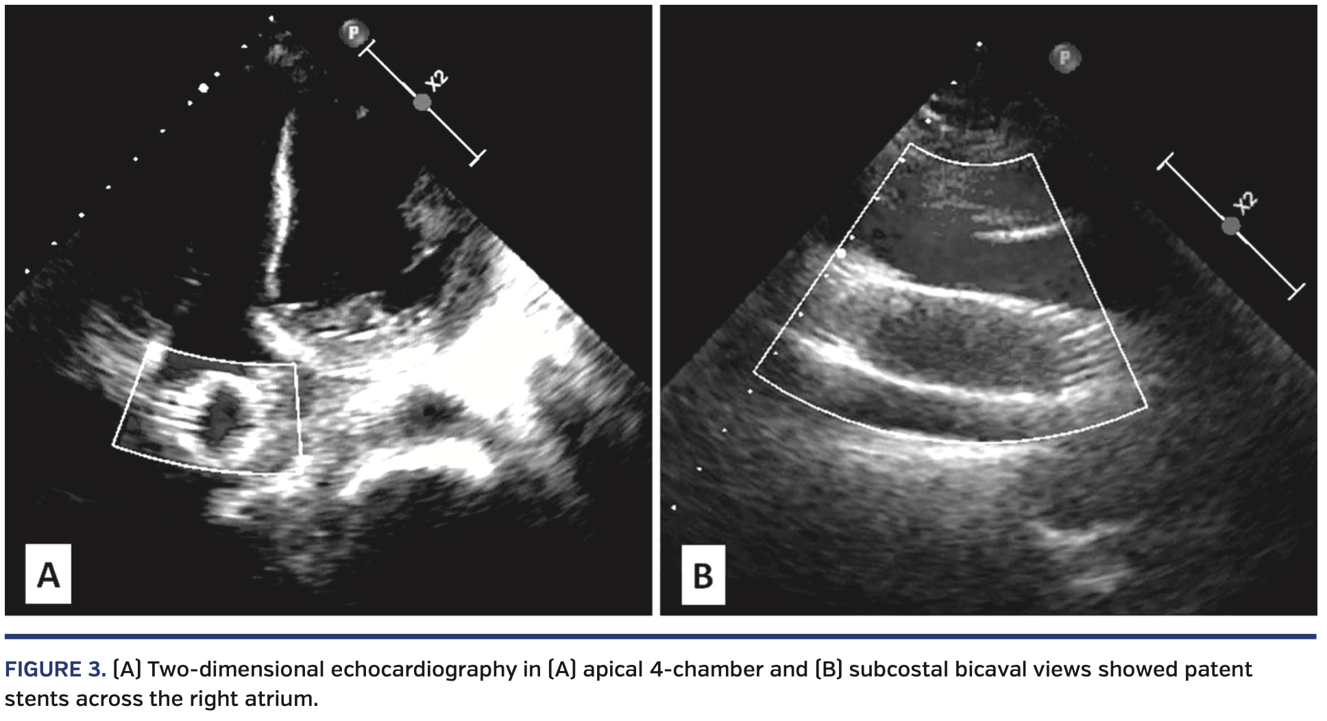
A 42-year-old female with end-stage chronic kidney disease, who was on maintenance hemodialysis for the last 3 years, presented with facial and upper-limb swelling of 2-month duration. As the 2-year-old right brachiocephalic fistula was not adequately functional, she was put on hemodialysis from the groin. Clinical examination revealed markedly engorged neck veins (Figure 1A). A computed tomography (CT) scan confirmed significant stenosis of the superior vena cava (SVC) (Figure 2A). She underwent percutaneous stenting of the SVC with a 16 x 60 mm self-expanding Wallstent (Boston Scientific) at an outside hospital. In view of persistent symptoms of SVC obstruction and migration of the Wallstent into the right atrium (Figure 2B), she was referred to our center for further management. Following discussion in a multidisciplinary meeting, it was proposed to perform a repeat endovascular intervention to relieve the SVC obstruction and manage the migrated stent. Following insertion of an 11 Fr right femoral venous sheath, a 0.032˝ hydrophilic wire (Terumo Medical) was placed in the SVC through the lumen of the migrated right atrial stent. The stenosed SVC segment was dilated with a 14 x 40 mm Atlas balloon (Bard Peripheral Vascular) (Figure 2C). A 24 x 100 mm self-expanding Sinus XL non-graft stent (OptiMed Medizinische Instrumente) was deployed, extending from the SVC to the inferior vena cava, through the migrated right atrial stent using the bridging-stent technique (Figures 2D and 2E). The SVC segment was further dilated with a 16 x 40 mm Atlas balloon. A brisk flow was achieved through the SVC (Video 1). Post intervention, hemodialysis could be restarted from the right forearm. At 6-month follow-up exam, she had marked improvement in her neck veins (Figure 1B), and two-dimensional echocardiography could delineate the well-positioned, tunneled stents across the right atrium (Figure 3).
From the Departments of 1Cardiology and 2Radiology, Advanced Cardiac Centre, Post Graduate Institute of Medical Education & Research, Chandigarh, India.
Disclosure: The authors have completed and returned the ICMJE Form for Disclosure of Potential Conflicts of Interest. The authors report no conflicts of interest regarding the content herein.
The authors report that patient consent was provided for publication of the images used herein.
Manuscript accepted August 20, 2019.
Address for correspondence: Prof (Dr) Rajesh Vijayvergiya, MD, DM, FSCAI, FISES, FACC, Department of Cardiology, Advanced Cardiac Centre, Post Graduate Institute of Medical Education & Research, Sector 12, Chandigarh–160 012, India. Email: rajeshvijay999@hotmail.com


