Heterotopic Ossification: A Late Complication From a Chemical Burn
Case Description
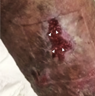
A 70-year-old man with a history of napalm burn to the posterior torso that he sustained while serving in the military during the Vietnam War presented with a firm mass on his right flank. Three years prior to this presentation, he developed a similar lesion on his left flank that was excised, with pathology confirming a diagnosis of heterotopic ossification (HO).
 |
| Figure 1. |
Questions
1. What is heterotopic ossification?
2. What causes heterotopic ossification?
3. What is the standard method of diagnosis?
4. What is the preferred management of heterotopic ossification?
Heterotopic Ossification
1. What is heterotopic ossification?
Heterotopic ossification is a relatively uncommon and debilitating complication associated with burns, spinal cord/neurologic injury, musculoskeletal trauma, and orthopedic surgery.1 It is defined as ectopic production of mature bone in nonskeletal tissue.2 Heterotopic ossification can occur anywhere on the body but has commonly been described as occurring on the upper extremities and overlying the hips. Cutaneous HO is largely asymptomatic; however, joint involvement often presents with pain and limitations in range of motion secondary to ankylosis. Interestingly, HO that occurs following burn injuries is not always limited to the site of cutaneous injury.3
Causes
2. What causes heterotopic ossification?
While the exact cause of HO is poorly defined, several research studies highlight the inflammatory response as well as infectious causes as precipitators for the disease. Studies found that the severity and incidence of HO is directly correlated to the degree of injury and inflammatory response of the inciting event.1 Early inflammatory markers, such as interleukin (IL)-3, IL-12, and IL-13, have been found to be associated with the development of HO when studied in combat-related blast injuries.4 These cytokines play important roles in lymphocyte differentiation as well as bone homeostasis through inhibiting osteoclastic activity by upregulating osteoprotegerin.1,5 As such, current research is investigating the role of the adaptive immune response in the development of HO.
Method of Diagnosis
3. What is the standard method of diagnosis?
Much attention has been paid in predicting the development of HO in patients with spinal cord injury and in those with combat-related injuries. Ultrasonography has been shown to be a reliable and highly sensitive screening modality for the diagnosis of HO in this patient population, with a sensitivity of 88.9%.6 The most sensitive imaging modality for early detection and for assessing the maturity of HO is a 3-phase technetium-99m (99mTc) methylene diphosphonate bone scan.2 The diagnosis of HO can be confirmed with magnetic resonance imaging or computed tomography.7
Preferred Management
4. What is the preferred management of heterotopic ossification?
Physical therapy interventions are aimed at maintaining joint mobility but are controversial in HO prevention and treatment, as various studies have shown that passive stretching has been associated with progression of ossification, eventually leading to complete ankylosis. However, other studies have found that daily aggressive stretch exercises have caused significant improvement in joint motion and can eliminate the need for further surgery.8 In addition to physical therapy interventions, indomethacin is frequently used in the prophylaxis and early treatment of HO due to its ability to prevent the inflammatory response that has been associated with HO development. Radiation therapy has been shown to be effective in the prophylaxis and prevention of progression of HO as a result of inhibition of mesenchymal cell differentiation. Pulse low-intensity electromagnetic field therapy, which utilizes magnetic fields to increase oxygen levels and decrease toxic by-products of inflammation by increasing local blood flow, has been shown to be effective in preventing the development of HO. Bisphosphonates have been shown to halt the progression of HO due to their role in preventing bone mineralization. Surgical excision should be delayed 12 to 18 months after development of HO until radiographic evidence of HO maturation. In addition, surgical excision should be supplemented with bisphosphonate therapy to prevent secondary HO formation.9
Conclusion
Our patient had a history of napalm burn to the posterior torso sustained in combat with the eventual development of HO on his left flank, which was excised and subsequently developed HO on the right flank 3 years later. Of note, he did not receive supplemental treatment with the aforementioned nonsurgical therapies, which may have prevented his second episode of HO.
Acknowledgments
Authors: Eric Clayman, MS,a Bahar Abbassi, MD,b Anthony W. Watt, MD,b and Wyatt G. Payne, MDb,c
Affiliations: aUniversity of South Florida Morsani College of Medicine, Tampa; bDivision of Plastic Surgery, Department of Surgery, University of South Florida Morsani College of Medicine, Tampa; and cC. W. Bill Young Bay Pines VA Medical Center, Bay Pines, Fla
Correspondence: abbassi@health.usf.edu
Disclosures: The authors disclose no financial or other conflicts of interest.
References
|
1. Ranganathan K, Agarwal S, Cholok D, et al. The role of the adaptive immune system in burn-induced heterotopic ossification and mesenchymal cell osteogenic differentiation. J Surg Res. 2016;206(1):53-61. |
|
2. Mavrogenis AF, Soucacos PN, Papagelopoulos PJ. Heterotopic ossification revisited. Orthopedics. 2011;34:177. |
|
3. Levi B, Jayakumar P, Giladi A, et al. Risk factors for the development of heterotopic ossification in seriously burned adults: a NIDRR burn model system database analysis. J Trauma Acute Care Surg. 2015;79(5):870-6. |
|
4. Forsberg JA, Potter BK, Polfer EM, et al. Do inflammatory markers portend heterotopic ossification and wound failure in combat wounds? Clin Orthop Relat Res. 2014;472:2845-54. |
|
5. Stein NC, Kreutzmann C, Zimmerman SP. Interleukin-4 and interleukin-13 stimulate the osteoclast inhibitor osteoprotegerin by human endothelial cells through the STAT6 pathway. J Bone Miner Res. 2008;23:750-8. |
|
6. Rosteius T, Suero EM, Grasmücke D, et al. The sensitivity of ultrasound screening examination in detecting heterotopic ossification following spinal cord injury. Spinal Cord. 2017;55(1):71-3. |
|
7. Ohlmeier M, Suero EM, Aach M, et al. Muscle localization of heterotopic ossification following spinal cord injury. Spine J. 2017;17:1519-22. |
|
8. Coons D, Godleski M. Range of motion exercises in the setting of burn-associated heterotopic ossification at the elbow: case series and discussion. Burns. 2013;39:e34-8. |
|
9. Teasell RW, Mehta S, Aubut JL, et al. A systematic review of the therapeutic interventions for heterotopic ossification after spinal cord injury. Spinal Cord. 2010;48:512-21. |


