Reconstruction of Oncologic Axillary Defects
Questions
1. What are the common indications and goals for axillary reconstruction?
2. What are the most commonly used flaps used in axillary reconstruction?
3. What are the most important postoperative considerations for axillary reconstruction?
4. What are the potential postoperative complications with axillary reconstruction?
Case Description
Case 1
A 52-year-old male was diagnosed with malignant melanoma of the axilla involving the axillary lymph nodes (Figure 1A). His disease progressed on immunotherapy, and he therefore underwent wide local excision of melanoma and axillary lymphadenectomy. This left a 13 x 8-cm defect (Figure 1B). Reconstruction was performed with a superiorly based transposition flap (Figures 1C and 1D).

Case 2
A 65-year-old female was diagnosed with right breast cancer for which she underwent lumpectomy and axillary lymph node dissection followed by radiation and chemotherapy. She developed an axillary recurrence (Figure 2A) for which she underwent axillary lymph node dissection and excision of overlying involved skin. This left a 15 x 8-cm cutaneous defect (Figure 2B). Reconstruction was performed with a thoracodorsal artery perforator flap (Figures 2C and 2D).

Case 3
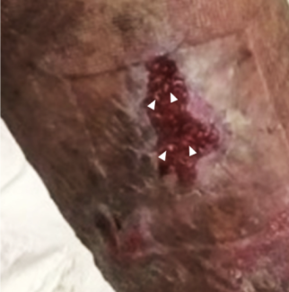
A 64-year-old female developed a fungating tumor in the left axilla. Biopsy and workup revealed a Merkel cell carcinoma with axillary lymph node involvement. The tumor did not respond to radiation and immunotherapy. Surgical resection, which included axillary lymph nodes, overlying skin, and parts of the pectoralis major and minor muscle, was performed (Figure 3A). This left a deep wound that was 15 x 8.5 cm in size. Reconstruction was performed with an anterolateral thigh free flap (Figure 3B). Microvascular anastomosis was performed to the thoracoacromial vessels.

Q1: What are the common indications and goals for axillary reconstruction?
The principal goals of axillary reconstruction are restoration of shoulder mobility by minimizing secondary contracture and coverage of neurovascular structures. The most common reasons for reconstruction are burn contractures, tumors, and hidradenitis suppurativa.1-4 Axillary burn contractures are seen most commonly in developing countries due to paucity of burn care and rehabilitation. Tumors of the axilla are most commonly breast cancers and cutaneous tumors with axillary lymph node involvement. Some oncologic defects may be extensive, requiring chest wall reconstruction. Hidradenitis suppurativa is a chronic recurring skin condition commonly involving the axilla. Management options consist of antibiotics, corticosteroids, immunosuppressants and biologic therapy (ie, adalimumab or infliximab), and surgical excision.5 Surgical options range from incision and drainage to localized or extensive resection.6
Q2: What are the most commonly used flaps in axillary reconstruction?
Axillary reconstruction can be performed with local flaps, regional pedicled flaps, and free flaps. Local flaps are used for smaller defects. Flap designs include transposition flaps, VY flaps, and rhomboid flaps.7,8 Defects that are large or require coverage of neurovascular structures may require regional pedicled flaps like the parascapular flap, latissimus dorsi myocutaneous flap, or pectoralis major muscle/myocutaneous flap.9 Regional perforator flaps decrease donor site morbidity by sparing muscle and allow for longer vascular pedicles. The thoracodorsal artery perforator flap is in an ideal location for axillary resurfacing.10 Free flaps are needed when defects are very large or when regional flap options are not available; tumor resection may include regional flap pedicles, and previous radiation may render regional tissue quality unsuitable for reconstruction. Common recipient vessels for free tissue transfer are the thoracodorsal and thoracoacromial vessels.7,11
Q3: What are important postoperative considerations for axillary reconstruction?
The most important consideration in the immediate postoperative period is to prevent compression of the flap or its vascular pedicle. As per our protocol, patients are kept in a 45-degree abduction splint. This is to prevent compression of the flap pedicle while not putting too much stress on the suture lines. After 2 weeks, abduction is gradually increased. Full movements without restrictions are allowed at 4 weeks. Patients need to be followed closely, as early aggressive shoulder motion may cause flap dehiscence, and prolonged immobilization can cause permanent shoulder stiffness.
Q4: What are the potential postoperative complications with axillary reconstruction?
Axillary reconstruction with flaps carries a risk of infection, bleeding, wound dehiscence, and flap necrosis.12 Hematoma may compress flap pedicle and axillary vessels. Lymphatic fluid leak from transected lymphatic vessels may cause prolonged drain output. Chyle leak is a rare complication that occurs due to transection of aberrantly terminating thoracic duct.13 Removal of axillary lymph nodes can lead to long-term upper extremity lymphedema. A recent meta-analysis found the risk of lymphedema after axillary lymph node dissection for breast cancer to be 24%.14 Prophylactic lymphovenous bypass at the time of lymph node removal is a newer technique that has been shown to decrease the incidence of lymphedema.15
Acknowledgments
Affiliations: Plastic and Reconstructive Surgery Service, Memorial Sloan-Kettering Cancer Center, New York, New York
Correspondence: Farooq Shahzad, MBBS, MS, FACS, FAAP; shahzadf@mskcc.org
Funding: This research was funded in part through National Institutes of Health/National Cancer Institute Cancer Center Support Grant P30 CA008748.
Disclosures: The authors disclose no financial or other conflicts of interest.
References
1. Geh JL, Niranjan NS. Perforator-based fasciocutaneous island flaps for the reconstruction of axillary defects following excision of hidradenitis suppurativa. Br J Plast Surg. 2002;55(2):124-128. doi:10.1054/bjps.2001.3783
2. Karki D, Ahuja RB. A review and critical appraisal of central axis flaps in axillary and elbow contractures. Burns Trauma. 2017;5:13. Published 2017 May 4. doi:10.1186/s41038-017-0079-7
3. Kim DC, Wright SA, Morris RF, Demos J. Management of axillary burn contractures. Tech Hand Up Extrem Surg. 2007;11(3):204-208. doi:10.1097/bth.0b013e318155946e
4. Miyamoto S, Fujiki M, Kawai A, Chuman H, Sakuraba M. Anterolateral thigh flap for axillary reconstruction after sarcoma resection. Microsurgery. 2016;36(5):378-383. doi:10.1002/micr.22529
5. Saunte DML, Jemec GBE. Hidradenitis suppurativa: Advances in diagnosis and treatment. Jama. 2017;318(20):2019-2032. doi:10.1001/jama.2017.16691
6. Vaillant C, Berkane Y, Lupon E, et al. Outcomes and reliability of perforator flaps in the reconstruction of hidradenitis suppurativa defects: A systemic review and meta-analysis. J Clin Med. 2022;11(19):5813. Published 2022 Sep 30. doi:10.3390/jcm11195813
7. Hallock GG. Island thoracodorsal artery perforator-based V-Y advancement flap after radical excision of axillary hidradenitis. Ann Plast Surg. 2013;71(4):390-393. doi:10.1097/SAP.0b013e31824b3e42
8. Netscher DT, Baumholtz MA, Bullocks J. Chest reconstruction: II. Regional reconstruction of chest wall wounds that do not affect respiratory function (axilla, posterolateral chest, and posterior trunk). Plast Reconstr Surg. 2009;124(6):427e-435e. doi:10.1097/PRS.0b013e3181bf8323
9. Amendola F, Cottone G, Alessandri-Bonetti M, et al. Reconstruction of the axillary region after excision of hidradenitis suppurativa: A systematic review. Indian J Plast Surg. 2023;56(1):6-12. Published 2022 Dec 11. doi:10.1055/s-0042-1758452
10. La Padula S, Pensato R, Pizza C, et al. The thoracodorsal artery perforator (TDAP) flap for the treatment of hidradenitis suppurativa of the axilla: A prospective Comparative Study. Plast Reconstr Surg. 2023;152(5):1105-1116. doi:10.1097/PRS.0000000000010435
11. Chen HC, Wu KP, Yen CI, et al. Anterolateral thigh flap for reconstruction in postburn axillary contractures. Ann Plast Surg. 2017;79(2):139-144. doi:10.1097/SAP.0000000000001097
12. Myers B. Understanding flap necrosis. Plast Reconstr Surg. 1986;78(6):813-814. doi:10.1097/00006534-198678060-00018
13. Chow WT, Rozen WM, Patel NG, Ramakrishnan VV. Chyle leak after axillary lymph node dissection. J Plast Reconstr Aesthet Surg. 2015;68(5):e105-106. doi:10.1016/j.bjps.2015.01.019
14. Che Bakri NA, Kwasnicki RM, Khan N, et al. Impact of axillary lymph node dissection and sentinel lymph node biopsy on upper limb morbidity in breast cancer patients: A systematic review and meta-analysis. Ann Surg. 2023;277(4):572-580. doi:10.1097/SLA.0000000000005671
15. Jorgensen MG, Toyserkani NM, Sørensen JA. The effect of prophylactic lymphovenous anastomosis and shunts for preventing cancer-related lymphedema: a systematic review and meta-analysis. Microsurgery. 2018;38(5):576-585. doi:10.1002/micr.30180


