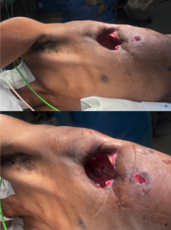
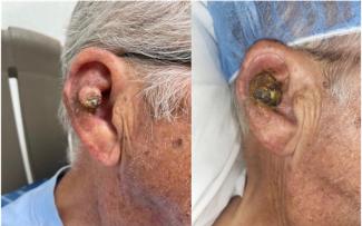
Tissue Is the Issue: Use of 2 Bipedicled “Bucket-Handle” Local Advancement Flaps to Close a Nonhealing Wound
Abstract
Introduction. Soft tissue loss following total knee arthroplasty can result in catastrophic complications. Defects can be covered using various flaps and grafts, including fasciocutaneous flaps. Here, we discuss one case of double bipedicled “bucket-handle” local advancement flaps used for a nonhealing midline knee dehiscence wound following total knee arthroplasty.
Methods. Flaps were planned using perforators identified with forward-looking infrared (FLIR) thermal imaging. Two bucket-handle bipedicled flaps were used for repair. Autologous split-thickness skin grafts were used for the donor sites.
Results. FLIR imaging was used for flap monitoring. Apart from one site of superficial epidermolysis that healed with local wound care, there were no postoperative complications.
Discussion. This case demonstrates the successful use of double bipedicled local advancement flaps to reconstruct a defect following a total knee arthroplasty. These flaps minimize donor site morbidity, provide adequate coverage, allow for tension-free closures, and have reliable vascular supplies. FLIR thermal imaging is an accessible and useful tool in designing and monitoring flaps.
Introduction
Soft tissue defects following total knee arthroplasty (TKA) are debilitating complications that can have devastating outcomes, including removal of the prosthesis or amputation.1 Risk factors for wound healing failure include smoking and diabetes. Defect coverage following TKA can be done in several ways, each with its own indications and potential complications. Skin grafts may be indicated in the case of shallow defects but are not commonly used because the graft contracture may compromise mobility of the knee, and failure of the graft is likely to result in an exposed prosthesis.2 Muscular and musculocutaneous flaps are indicated for larger defects but may result in higher donor site morbidity than other forms of reconstruction.2 A third useful flap, first described in 1981 by Pontén, is the fasciocutaneous flap.3
The fasciocutaneous flap involves the skin, subcutaneous tissue, and fascia and has enhanced vascularity due to the underlying fascial plexus.4 Pontén first demonstrated its safe use in lower leg reconstruction with length:width ratios of up to 3:1.3 This flap can be designed in multiple ways, including as a free flap, a regional flap, and a local advancement flap.5
The local advancement flap limits donor site morbidity and preserves musculature to maximize postoperative lower extremity mobility, while reserving potential flaps for future reconstructive needs.6 Using multiple pedicles instead of a single pedicle can reduce morbidity by augmenting vascular supply. Here, we report one case of double bipedicled “bucket-handle” local advancement flaps for closure of a midline, nonhealing surgical dehiscence wound following a TKA revision.

Methods
An 81-year-old man with a history of right-sided TKA in 2016 and revision in June 2022 was admitted from the wound care clinic with a nonhealing surgical dehiscence wound draining increasingly copious serous fluid (Figure 1). The patient was initially treated with a 6-week course of amoxicillin-clavulanate, vancomycin, and piperacillin-tazobactam, which failed to resolve the infection. Preoperative exploration revealed a tunneled wound communicating with the underlying TKA at the site of the lateral capsulotomy incision, and flap closure was planned. Intraoperatively, a full-thickness wound was discovered at the lateral capsulotomy site with a 6 × 4-cm area of skin necrosis along the midline incision. Wound cultures were obtained, and the joint space and prosthesis were irrigated and debrided. Acellular dermal matrix (ADM) was placed to cover the joint arthroplasty, and the patellar tendon was imbricated by the orthopedic surgeon. At this stage, an 8 × 6-cm region of ADM and a 7 × 7-cm region of patellar tendon were exposed (Figure 2).

The wound required medial and lateral soft tissue coverage, so a local advancement bipedicled fasciocutaneous flap from the medial aspect of the knee was first performed. Intraoperatively, forward-looking infrared (FLIR) thermal imaging was used to identify perforators by examining the temperature of various sites along the proposed flap (Figure 3A, 3B). Arterial perforators are higher in temperature and appear whiter in color with FLIR imaging, providing a visual guide. A 7 cm-wide flap was delineated (Figure 3A, 3B). Sharp subcutaneous dissection was performed, and the flap was advanced laterally 4 cm. To cover the remaining defect, a lateral flap was done in the same fashion, using a 7 cm-wide flap with perforators again identified using FLIR. This provided adequate coverage for the ADM and tendon without exposing the prosthesis.

To close the 2 donor site defects, the posterior soft tissue from the medial and lateral knee was advanced anteriorly and sutured to the wound base with absorbable sutures, reducing the defect sizes to 16 × 2 cm and 17 × 2 cm. These were covered with autologous split-thickness skin grafts. The midline incision had a 15 × 3-mm region of superficial epidermolysis near the original site of wound dehiscence. An extracellular matrix graft and silver sulphate antimicrobial dressing were used to cover the epidermolysis as well as the skin grafts.
Results
Incisional negative pressure wound therapy was started in the operating room and continued for 10 days, and a knee immobilizer was placed to prevent shearing of the flaps. The patient was admitted to the hospital for 1 week before being transferred to a long-term acute care hospital. Mobilization began on postoperative day 1 with a rolling walker, taking care to avoid weight-bearing on the right lower extremity.

Infectious disease was consulted to determine the best course of antibiotic therapy and continued to follow him during his stay in the long-term acute care facility and after his discharge. He received 6 weeks of intravenous vancomycin and cefepime for hardware infection and osteomyelitis. Cefepime was chosen instead of piperacillin-tazobactam to reduce risk for acute kidney injury. Following completion of intravenous antibiotics, the patient began oral suppressive therapy with doxycycline. All intraoperative wound cultures were negative for growth; however, the patient did receive preoperative antibiotic therapy. Pain management was with acetaminophen, naproxen, and oxycodone as needed, although after the first week, the patient’s pain was well-controlled with acetaminophen and naproxen. Postoperative flap viability was monitored using FLIR thermal camera and visual inspection (Figure 4A, 4B). Figure 4A and Figure 4B demonstrate flap monitoring performed during the first 24 hours. Despite some postoperative skin discoloration seen on visual inspection, both the medial and lateral flaps were well-perfused, with no regions of deep red or violet on FLIR imaging.

The patient spent 6 weeks at a long-term acute care hospital where he completed physical therapy for gait training, reducing edema, and improving range of motion, per the total knee protocol. At his 12-week postoperative follow-up appointment, radiologic studies demonstrated anatomic alignment of the right knee without any loosening. On physical exam, the patient had 0 to 110 degrees of flexion at the knee and ambulated with a normal gait. There were no signs of inflammation or infection.

The patient had no postoperative complications except for a 0.6 × 0.6-cm area of superficial epidermolysis that healed uneventfully with dressing changes every 3 days. His 2-week postoperative photographs are shown in Figure 5 and Figure 6. The epidermolysis resolved completely (Figure 5), and the skin graft recipient site also healed without incident (Figure 6). The small sites of necrosis along the graft recipient site were managed with 10 days of silver sulphate antimicrobial dressing followed by foam border dressings; these sites were healed by 3 weeks after surgery. At his most recent follow-up visit, the patient had regained near full functionality of the affected limb.
Discussion
This report demonstrates the successful use of the double bucket-handle local advancement flap for closure of a midline knee defect following TKA incision dehiscence, as well as the utility of FLIR thermal imaging for surgical planning and flap monitoring.
Although the bilateral gastrocnemius flap was initially considered, the flap dimensions did not provide adequate coverage and we sought to use a muscle-sparing option to preserve function. In patients with a total knee replacement, mobility is already a challenge, and muscle-based flaps can make recovery more difficult. Several potential types of flaps maximize postoperative functionality. A fasciocutaneous flap such as this one preserves all musculature, while a free muscle transfer flap like the rectus abdominis preserves leg muscle. Compared with free flaps, however, local fasciocutaneous flaps are simpler to execute, require shorter operative time, have better skin color and quality match, and offer less donor site morbidity.7
For this surgery, FLIR imaging was used to identify suitable perforators to design the flap and to monitor intraoperative and postoperative vascularity. Thermal imaging uses temperature changes as a surrogate measure of vascularity, providing a noninvasive estimate of flap blood supply. A yellow or white color suggests higher temperature and corresponds with increased perfusion. On the other hand, a blue or violet color is associated with lower temperatures, suggesting reduced perfusion.8 Prior studies have demonstrated that FLIR has a high positive predictive value (97.4%) and negative predictive value (85.7%) for flap failure.9 This present case report corroborates previous evidence and demonstrates its usefulness not only for flap monitoring but also for surgical planning. In cases where such tools as fluorescent angiography imaging are unavailable, FLIR is an accessible alternative.
Fasciocutaneous flaps can be raised in several ways. For example, a single pedicle can be used, based on vessels including the sural artery or anterior tibial artery.2 However, these may have a less reliable vascular supply than bipedicled flaps, where the additional vascularity reduces risk of flap loss. As seen in this case, using 2 bipedicled flaps has the added advantage of reducing tension on the closure, thus improving flap survival and allowing for coverage of larger defects.
The double bucket-handle flap has rarely been described in the literature for knee coverage,2,6 but its usefulness is multifold. As written by Batchvarova and Masquelet in 2007, these flaps are highly reliable due to a rich anastomotic network around the knee, consisting of 3 vessels (the descending genicular, lateral femoral circumflex, and popliteal arteries) connected by several lateral genicular arteries.2 Flap mobilization requires division of the horizontal vascular supply, but this in turn induces increased vascularity of the remaining vessels. Additionally, perforators can be preserved so long as they do not restrict mobilization of the flap, further augmenting the vascular supply.2
Some patients may need to return to the operating room due to flap failure or other complications. In these patients, starting with a more conservative choice for coverage, like a local flap, may help preserve potential future options.
One drawback of this option for reconstruction is the creation of bilateral donor sites that must be covered. We elected to close these donor sites with split-thickness skin grafts to decrease healing time. This step does require additional operative time and a graft donor site, but usage of the anterolateral thigh ensures good match of skin color and quality.
The double bucket-handle local advancement flap is an underutilized flap in closure of midline knee defects. This case report adds to the literature to support its increased use. FLIR thermal imaging is an innovative method of flap planning and monitoring that is accessible, noninvasive, and accurate and helps to ensure improved success of these local flaps.
Acknowledgments
Affiliations: 1Tulane University School of Medicine, New Orleans, Louisiana; 2Department of Surgery, Tulane University School of Medicine, New Orleans, Louisiana; 3Division of Plastic and Reconstructive Surgery, Department of Surgery, Tulane University School of Medicine, New Orleans, Louisiana
Correspondence: Abigail E Chaffin, MD; achaffin@tulane.edu
Disclosures: The authors disclose no relevant conflicts of interest or financial disclosures for this manuscript.
References
1. Laing JH, Hancock K, Harrison DH. The exposed total knee replacement prosthesis: a new classification and treatment algorithm. Br J Plast Surg. 1992;45(1):66-69. doi:10.1016/0007-1226(92)90120-m
2. Batchvarova Z, Masquelet AC. Bipedicled fasciocutaneous flaps for coverage of defects of the knee [French]. Ann Chir Plast Esthet. 2007;52(2):124-129. doi:10.1016/j.anplas.2006.05.002
3. Pontén B. The fasciocutaneous flap: its use in soft tissue defects of the lower leg. Br J Plast Surg. 1981;34(2):215-220. doi:10.1016/s0007-1226(81)80097-5
4. Haertsch P. The surgical plane in the leg. Br J Plast Surg. 1981;34(4):464-469. doi:10.1016/0007-1226(81)90060-6
5. Cormack GC, Lamberty BGH. The arterial anatomy of skin flaps. 2nd ed. Churchill Livingstone; 1994.
6. Granzow JW, Li A, Suliman A, Caton A, Goldberg M, Boyd JB. Bipedicled flaps in posttraumatic lower-extremity reconstruction. J Plast Reconstr Aesthet Surg. 2013;66(10):1415-1420. doi:10.1016/j.bjps.2013.05.019
7. Jordan DJ, Malahias M, Hindocha S, Juma A. Flap decisions and options in soft tissue coverage of the lower limb. Open Orthop J. 2014;8:423-432. doi:10.2174/1874325001408010423
8. Paul SP. Using a thermal imaging camera to locate perforators on the lower limb. Arch Plast Surg. 2017;44(3):243-247. doi:10.5999/aps.2017.44.3.243
9. Rabbani MJ, Bhatti AZ, Shahzad A. Flap monitoring using thermal imaging camera: a contactless method. J Coll Physicians Surg Pak. 2021;30(6):703-706. doi:10.29271/jcpsp.2021.06.703


