The “Domino Flaps” Concept: Functional Composite Reconstruction for Traumatic Transmetacarpal Thumb Amputation
© 2023 HMP Global. All Rights Reserved.
Any views and opinions expressed are those of the author(s) and/or participants and do not necessarily reflect the views, policy, or position of ePlasty or HMP Global, their employees, and affiliates.
Abstract
Complex transmetacarpal thumb amputation remains a challenging reconstructive injury. Optimal reconstructive options aim to achieve a neo-thumb with optimal length, sensitivity, stability, and an aesthetically functional result. In cases when immediate replantation of the amputated digit is not possible, a temporary ectopic replantation with staged reconstruction can be deployed. We report our experience of a complex transmetacarpal thumb amputation managed with a staged “domino flap” concept. The first stage involved an ectopic replantation of the amputated digit with a second stage replantation 3 weeks later. Domino flap refers to the requirement of a further reconstruction due to the defect at the donor sites. In this case, the replant is accompanied by 2 domino flap reconstructions with the dorsalis pedis composite free flap to reconstruct the first metatarsal and an anterior tibial artery propeller perforator flap to reconstruct the composite flap donor site.
Introduction
Transmetacarpal thumb amputation is a complex traumatic condition. Treatment algorithms depend on the severity of the injury and include replantation, toe-to-thumb transfer, osteoplastic approaches, and other modified soft tissue reconstruction methods.1-3 The aim of reconstruction is to achieve a neo-thumb with an aesthetically functional result,3-5 but sometimes immediate replantation of the amputated digit is not possible. In those circumstances, the concept of temporary ectopic replantation has been described, with the first successful case being described by Godina (1986).6-8 For first metacarpal joint reconstruction following amputation, Guo-xian et al in 1995 described the use of the second metatarsal flap to restore the anatomy of the first metacarpophalangeal joint (MCPJ).4,8-10 When using this flap, it is important to also consider the donor site of the second metatarsal flap and determine appropriate soft tissue coverage. In this paper, we report our experience of a complex transmetacarpal thumb amputation that was managed with a staged “domino flap” concept. The first stage involved an ectopic replantation of the amputated digit with a second stage replantation 3 weeks later. Domino flap refers to the requirement of a further reconstruction due to the defect at the donor sites. In this case, the replant is accompanied by 2 domino flap reconstructions with the dorsalis pedis composite free flap to reconstruct the first metatarsal and an anterior tibial artery propeller perforator flap to reconstruct the composite flap donor site.
Case Report
A 31-year-old man with a severe transmetacarpal left thumb amputation presented to our department. The mechanism of injury included both crushing and shearing forces following entrapment of his hand in an industrial plastic press. On examination, he had sustained a transmetacarpal amputation of his left thumb with MCPJ and first metacarpal bone loss. Accompanying this was an injury to the tendons and an extensive area of skin and soft tissue damage. A thumb replantation in this case carried a very high risk of failure due to the shearing and soft tissue damage, but the patient requested for the digit to be salvaged. Therefore, we opted for a temporary ectopic replantation of the amputated thumb to the lateral thigh. Three weeks later, the ectopic implanted thumb was transferred from the thigh to its original anatomical position using a microvascular anastomosis. A composite vascularized second metatarsal osteotendinous flap was utilized to restore the anatomy of the crushed first metacarpal. Finally, the metatarsal donor site was repaired with iliac crest bone and anterior tibial artery propeller perforator flap. Split-thickness skin grafting was used to cover the propeller flap donor site. The main aim of this reconstructive technique was to restore the function and cosmetic appearance of the thumb.
Operative Technique
The patient underwent an emergency exploration and debridement of the wound under general anesthesia. There was a complete amputation at the level of the MCPJ of the left thumb. This was accompanied by a crush injury to surrounding tissues extending from the site of amputation to the wrist, resulting in necrotic skin and soft tissue. The carpometacarpal joint (CMCJ), trapezoid bone, and muscles within the zone of injury were crushed and torn. Most superficial flexors and extensors were absent, nerves were avulsed, and both radial and ulnar digital arteries were absent. Although the MCPJ was not salvageable, the distal stump of the thumb was well preserved. Taking into consideration the patient’s wish to preserve his thumb, we decided to salvage the amputated thumb with temporary ectopic implantation.


A two-team approach was implemented to undertake the debridement. The first team performed the debridement and washout of the wound. They used a local perforator-based propeller (dorso-ulnar) flap to cover the defect as it offered immediate and easy coverage. The second team performed an ectopic implantation of the amputated thumb and anastomosed the vastus lateralis arterial muscle perforator branch to the ulnar digital artery of the thumb. Additionally, the 2 accompanying veins of the vastus lateralis artery were anastomosed to the 2 dorsal thumb veins (Figure 1A). By the end of the procedure, the implanted thumb was well perfused (Figure 1B). The total ischemia time from the time of injury to the reperfusion was 5 hours.
Three weeks later, the ectopic thumb and flap had survived, and the second stage of the procedure was carried out to replant the thumb to its original anatomic position.


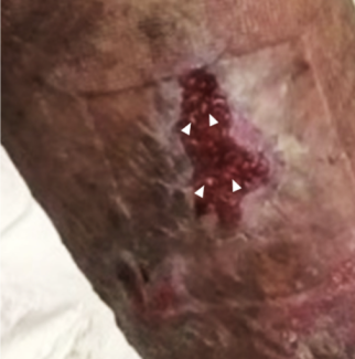
Before implanting the ectopic thumb (Figure 2), a composite dorsalis pedis flap was used to reconstruct the first MCPJ, flexor and extensor tendons, and neurovascular bundles (Figure 3). The composite flap donor site was reconstructed with iliac crest bone graft and anterior tibial artery propeller perforator flap (Figures 4, 5, and 6). The composite dorsalis pedis flap was fixed with a Kirschner wire, and the thumb was then transferred to its original anatomical site (Figure 7). A flexor hallucis longus and extensor digitorum tenorraphy was performed to the thumb flexor and extensor tendons respectively. The thumb ulnar digital artery was anastomosed to the dorsalis pedis artery, and the two veins of the radial artery were anastomosed to the veins of the dorsalis pedis artery. The ulnar digital nerve of the thumb was sutured to the to the superficial peroneal nerve (Figure 8). The propeller flap donor site was covered with a skin graft. Good blood supply was confirmed at the end of the thumb replantation procedure (Figure 9). This was dubbed the “domino” flap as it involves multiple donor and graft reconstructions.


Results
Postoperative recovery was uneventful, and early hand physiotherapy commenced 2 weeks after wound healing was achieved. In the 1-year follow-up, the thumb and thenar eminence had healed well with no concerns regarding wound healing nor scar appearance (Figure 10). Sensation was restored with light touch and 2-point discrimination being present. Active movements, such as MCPJ extension and a hook grip, were restored (Figure 11). A radiograph showed the first carpometacarpal joint in a good position with the fracture having healed without any bone resorption (Figure 12). The iliac crest bone graft used to reconstruct the second metatarsal had also healed well (Figure 13).
Discussion
A finger amputation combined with a large skin defect, soft tissue loss, CMC joint loss, and bone defect poses a significant challenge to the reconstructive surgeon. However, with the recent developments of microsurgical reconstruction techniques, ectopic limb replantation and foot composite tissue flap can provide an alternative solution so that amputated limbs can be retained and replanted.
Over the last few decades, successful finger, forearm, foot, and penis ectopic replantation have been reported.2-7 We believe that temporary ectopic replantation can be beneficial in cases of a proximal or distal amputated limb, particularly if there is a segmental injury where vascular anastomosis is not possible or significant tissue loss makes an emergency replantation extremely high risk. It can also be considered in patients with an amputation and concurrent vital organ damage unable to undergo a prolonged replantation procedure. The temporary ectopic replantation technique can be performed until the patient is clinically stable to undergo a limb replantation with repair of structures.
Ectopic foster replantation technique is currently only considered in a minority of cases, and some are still skeptical about the postoperative prognosis.12 When deciding to proceed with ectopic temporary replantation, clinicians must be aware of replantation risks, such as increased hospital stay, extensive surgery, and increased health care costs, and a relatively poor functional outcome may result. Therefore, before deciding to undertake this option, one must exclude all alternative options, such as replantation of a shortened limb, flap coverage, or amputation. However, taking into account the huge psychological impact to the patient, temporary foster care ectopic replantation may pose an invaluable option.1
Indications for ectopic limb replantation include patients with multiple injuries who cannot tolerate prolonged surgery, those with an ability to restore function after replantation, and those needing complex immediate replantation surgery requiring a warm ischemia time in excess of 10 hours. Some prerequisites to performing ectopic replantation include a healthy superficial vein in the uninjured limb, absence of chronic organic damage, and tolerance for multiple surgeries, and, most importantly, a strong wish for replantation. In the case described, the ectopic replantation method was chosen as this was a complex injury with an extensive soft tissue defect. The total warm ischemia time to perform the reconstruction as a single-stage procedure would be extremely long with negative impacts on the functional recovery, infection risks, tissue necrosis, and amputation likelihood.
Looking at areas suitable for ectopic replantation, Godina et al suggested that the thoracodorsal artery is the most suitable area. The thoracodorsal artery can be matched to the amputation site for the caliber of blood vessels.8 Graf et al suggested other options, such as the radial artery due to its large diameter, easy access, and its potential for enabling patients to use their own finger for passive activities.9 Other authors have chosen the groin area, using the inferior epigastric artery and saphenous vein as a retrograde inversion vein.9,10 Wang Jiangning et al discussed the use of contralateral limb as a foster region, describing such advantages as a large recipient vessel caliber and superficial location.11,12
In our case, we believe that despite the various options available for ectopic replantation of a digit, the anterolateral thigh region was the most suitable region. The advantages of this region are mobility of skin, a non-trunk vessel for anastomosis, and the easy access to care for the ectopic thumb before a second surgery.
Reported ectopic limb duration varies in the literature with the shortest being 26 days, the longest 156 days, and the average duration 42 days.13 However, the longer the ectopic replant duration, the worse the functional recovery after replantation.13 Therefore, provided that the general condition of the patient is stable, we are in favor of relatively early replantation to ensure a good recovery and limb function. In this case, as the wound appeared to be healing well 3 weeks following ectopic replantation, it was thought to be an appropriate time to carry out the anatomical site replantation.
The first metacarpal defect combined with a skin defect posed a significant reconstructive challenge; because of the whole segment defects, a composite second metatarsal flap was performed. Using a second metatarsal composite flap allowed the transfer of the toe joint as well as associated structures, such as tendons, vessels, and nerves.13 The composite flap ensures good blood circulation that allows for improved tissue remodeling and repair of the metacarpal hand wound; this flap also avoids joint and surrounding tissue degeneration, reduces the chance of scar adhesions, decreases pain, and improves the shape of hand injuries and joint function to improve functional outcomes.
The bone loss at the composite flap donor site was repaired with iliac crest bone graft to maintain the continuity and stability of the foot arch. An anterior tibial artery propeller flap was used to cover the soft tissue defect. A pedicled flap was chosen rather than a free flap as it provided a good functional and aesthetic result whilst minimizing the need for microvascular anastomoses.
Conclusions
In summary, we believe that the concept of “domino flaps” combined with ectopic replantation of the digit is a good option for reconstruction of what would otherwise be a nonreplantable digital amputation. We have shown that using this technique can yield both an aesthetically and functionally good outcome.
Acknowledgments
Authors DS, GP, and YS contributed equally to this manuscript.
Affiliations: 1Department of Oncology Plastic Surgery, Hunan Province Cancer Hospital, Hunan, China; 2Group for Academic Plastic Surgery, The Blizard Institute, Barts and The London School of Medicine and Dentistry, Barts Health NHS Trust, Queen Mary University of London, London, United Kingdom; 3Department of Reconstructive and Microsurgery, Lishui People’s Hospital, Zhejiang, China; 4University College London, London, United Kingdom; 5Plastic Surgery Department, Royal London Hospital, London, United Kingdom; 6Reconstructive Microsurgery Center, Suqian’s Third People’s Hospital, Jiangsu, China; 7The Third Department of Burns and Plastic Surgery and Center of Wound Repair, the Fourth Medical Center of PLA General Hospital, Beijing, China
Correspondence: Georgios Pafitanis, MD, PhD; georgios.pafitanis@nhs.net
Funding: Funding for this research was provided by Hunan Provincial Science Foundation grant (2018JJ2242), Hunan Provincial Science Foundation grant (2018JJ2241), Hunan Provincial science and technology planning project (2014SK3002), Hunan Provincial Health and Family Planning Commission project (B2014-111).
Disclosures: The authors disclose no relevant financial or nonfinancial interests.
References
1. Tomlinson JE, Hassan MSU, Kay SP. Temporary ectopic implantation of digits prior to reconstruction of a hand without metacarpals. J Plast Reconstr Aesthet Surg. 2007;60(7):856-860. doi:10.1016/j.bjps.2007.04.001
2. Graf P, Gröner R, Hörl W, Schaff JR, Biemer E. Temporary ectopic implantation for salvage of amputated digits. Br J Plast Surg. 1996;49:174-177. doi:10.1016/s0007-1226(96)90221-0
3. Yousif NJ, Dzwierzynski WW, Anderson RC, Matloub HS, Sanger JR. Complications and salvage of an ectopically replanted thumb. Plast Reconstr Surg. 1996;97:637-640. doi:10.1097/00006534-199603000-00024
4. Wang JN, Tong ZH, Zhang TH, et al. Salvage of amputated upper extremities with temporary ectopic implantation followed by replantation at a second stage. J Reconstr Microsurg. 2006;22:15-20. doi:10.1055/s-2006-931901
5. Matloub HS, Yousif NJ, Sanger JR. Temporary ectopic implantation of an amputated penis. Plast Reconstr Surg. 1994;93:408-412. doi:10.1097/00006534-199402000-00031
6. Ramdas S, Thomas A, Kumar SA. Temporary ectopic testicular replantation, refabrication and orthotopic transfer. Indian J Plast Surg. 2007;40:209-212. doi:10.1016/j.bjps.2007.01.074
7. Wang JN, Wang DM, Wang SY, et al. Temporary ectopic implantation for salvage of amputated lower extremities: case reports. Microsurgery. 2005;25:385-389. doi:10.1002/micr.20135
8. Godina M, Bajec J, Baraga A. Salvage of the mutilated upper extremity with temporary ectopic implantation of the undamaged part. Plast Reconstr Surg. 1986;78:295-299. doi:10.1097/00006534-198609000-00003
9. Kayikçioğlu A, Ağaoğlu G, Nasir S, Keçik A. Crossover replantation and fillet flap coverage of the stump after ectopic implantation: a case of bilateral leg amputation. Plast Reconstr Surg. 2000;106:868-873. doi:10.1097/00006534-200009020-00019
10. Chernofsky MA, Sauer PF. Temporary ectopic implantation. J Hand Surg Am. 1990;15:910-914. doi:10.1016/0363-5023(90)90014-i
11. Zhou MW, Li KD, Wang RJ, et al. Temporary ectopic implantation for salvage of amputated upper extremity. Chin J Hand Surg. 2001;17:67.
12. Tomlinson JE, Hassan MSU, Kay SP. Temporary ectopic implantation of digits prior to reconstruction of a hand without metacarpals. J Plast Reconstr Aesthet Surg. 2007;60(7):856-860. doi:10.1016/j.bjps.2007.04.001
13. Higgins J. Ectopic banking of amputated parts: A clinical review. J Pract Hand Surg. 2011; 36(11):1868-1876.


