Leukoplakia of the Lower Lip Reconstructed With a Tongue Flap
© 2024 HMP Global. All Rights Reserved.
Any views and opinions expressed are those of the author(s) and/or participants and do not necessarily reflect the views, policy, or position of ePlasty or HMP Global, their employees, and affiliates.
Questions
1. How is leukoplakia diagnosed?
2. How is leukoplakia managed and treated?
3. How is the lower lip reconstructed?
4. What is the purpose of a tongue flap?
Case Description
A 47-year-old man with a history of smoking tobacco presented with an ulcer on the lower lip persisting for 1 year. On examination, a white plaque (35 × 5 mm) was observed on the lower lip with no lesions on the lip mucosa (Figure 1). A biopsy was performed, and hematoxylin and eosin staining showed thickening of keratin with hyperkeratosis, intercellular edema, and prolonged protrusions in the epithelium. Immunohistochemical staining with Ki-67, p16, and p53 revealed the presence of dysplasia (Figure 2).

Figure 1. A homogeneous white plaque (35 × 5 mm) on the lower lip.

Figure 2. Histopathology of leukoplakia. (a) Hematoxylin and eosin staining (original magnification, ×100) showing keratin thickening with hyperkeratosis, intercellular edema, and prolonged protrusions in the epithelium. (b-d) Immunohistochemical staining; (b) Ki67, (c) p16, and (d) p53.
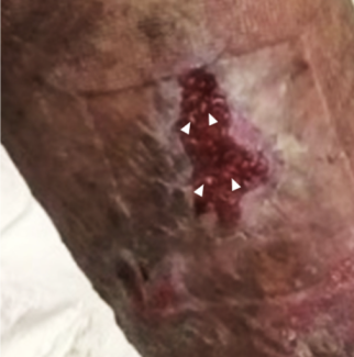
Based on these findings, leukoplakia was suspected. The lesion was resected with a 5-mm margin, and the defect covered two-thirds of the lower lip. Reconstruction of the lower lip was performed 3 weeks after the tumor resection. A tongue flap with a superior pedicle designed on the reverse side of the tongue tip was applied to the defect and sutured. Two weeks later, the tongue flap was divided, and the donor site was sutured (Figure 3). There was no recurrence during the 6-month follow-up period. The lip and tongue showed normal function, and the color and texture of the reconstructed lip also showed a good recovery. (Figure 4).

Figure 3. (a) Design of the resection with 5-mm margin. (b) The defect covering two-thirds of the lower lip. (c) The flap designed on the inferior surface of the tongue. (d) The flap harvested with a superior pedicle, 3-mm thick. (e) The flap applied over the defect. (f) The flap divided 2 weeks after the last operation.

Figure 4. The color and texture of the reconstructed lip (a) and the donor site (b) 6 months after the operation.
Q1. How is leukoplakia diagnosed?
Leukoplakia is defined by the World Health Organization (WHO) as a "white plaque of questionable risk having excluded [other] known diseases or disorders that carry no increased risk for cancer".1 The pooled prevalence estimated for leukoplakia of the oral cavity is between 1.5% and 2.6%, with no gender predilection;2 the mean age of patients with leukoplakia is between 50 and 60 years.3-4 Leukoplakia is more common on the lateral border of the tongue, buccal mucosa, and upper and lower gingiva/alveolar mucosa. The risk factors associated with oral leukoplakia include tobacco smoking, heavy alcohol consumption, betel nut chewing, and ultraviolet light exposure (for lesions of the lip).2
After excluding fungal infections using the KOH test, a biopsy is usually performed to confirm the diagnosis. Acanthosis, hyperkeratosis, irregular epithelial stratification, and loss of basal cell polarity are common pathological findings in patients with leukoplakia.4 Immunohistochemical staining (Ki-67, p16, and p53) is useful for revealing the presence of dysplasia.5 Most oral leukoplakias are benign, but some (0.13–36.4%) progress to oral squamous cell carcinoma.6 Therefore, a definitive surgical resection should be performed. The risk factors for malignant transformation are male sex, long duration, nonhomogeneous appearance, tongue/floor/soft palate location, size >200 mm, and dysplasia.6 However, the progression from dysplasia to invasive cancer is unclear.3
Q2. How is leukoplakia managed and treated?
Leukoplakia is a precancerous lesion with the potential for malignant transformation requiring definitive resection. Treatment methods include carbon dioxide lasers, photodynamic therapy, and medical treatment (ie, use of topical retinoid, 0.5–1% bleomycin, cyclooxygenase inhibitors, and phytochemical-enriched agents); however, these are not radical cures.2 Recurrence rate after surgical resection has been reported to range from 0% to 35%.2 Vedtofte et al reported that leukoplakia resected with a 3 to 5-mm margin recurred in 20% of patients because the margins of nonhomogeneous lesions are particularly difficult to precisely determine.7 Kuribayashi et al performed a Lugol iodine staining for identifying dysplastic epithelial tissues and suggested that an adequate resection margin (>3 mm) may reduce the risk of recurrence, irrespective of dysplasia grade.3 For less demarcated lesions, a margin of 5 mm can be taken where anatomically possible tissue is excised down to superficial striated muscle or periosteum with a depth of 3 to 5 mm. In the case of this patient with unclear demarcation of lesion, a resection with a 5-mm margin was performed and no lesions were found at the pathologic margins. No recurrence was observed during the 6-month follow-up period.
Q3. How is the lower lip reconstructed?
An important aspect of the lower lip reconstruction is that it is noninvasive and does not affect function, and the outcome is satisfactory in terms of color and texture. The lower lip requires reconstruction when the defects occupy more than one-third of the area. For superficial defect reconstruction, mucosal grafts, retrolabial mucosal advancement flaps (retrolabial flaps), and tongue flaps are often selected.8-9
The mucosal grafts have the advantage of fewer donor sacrifices; however, complications such as postoperative shrinking and deformity may occur.8 Ito et al reported the use of a hard palate mucoperiosteal graft to successfully reduce postoperative shrinking.8 This graft is useful for managing partial defects; however, it is difficult to use for long horizontal defects because the amount of grafts that can be harvested is limited. The most appropriate site for graft harvesting is the canine-premolar area 8 to 13 mm from the mid-palatal aspect of each respective tooth.10 Although there are individual differences in the limits of the size of the graft that can be harvested, it is often used for defects of about 20 × 10 mm. 8,10,11
The retrolabial flap technique is used to advance the posterior mucosa of the lower lip.9 This technique is very simple and often used; however, postoperative shrinkage may occur, and the vermillion area is pulled back when the coverage area is large.
The tongue flap has rich blood flow, and reconstruction using this flap is simple. Postoperative shrinkage occurs less frequently with a tongue flap than with a mucosal graft or a retrolabial flap. Here, the tongue flap technique was selected because of the anticipated defect and patient age.
Q4. What is the purpose of a tongue flap?
Lexer first used a tongue flap for vermilion reconstruction in 1909.12 The tongue flap has rich blood flow, a relatively wide area of mucosa, and greater strength and flexibility than those of other areas of the oral mucosa. This technique is very simple; however, it requires 2 operations for suturing and separation, and patients are restricted from eating and tongue movements until the flap is divided. Therefore, this flap cannot be used in patients with dysphagia or compliance problems, and identifying suitable patients is necessary. Vermilion reconstruction with a tongue flap is a good indication for resection of leukoplakia, given the superficial, long horizontal defect and the favorable age of onset. The above described patient met the above criteria, and the patient was on a liquid or soft diet in the hospital for 2 weeks after suturing the tongue flap to maintain rest and cleanliness in the region.
The key points of tongue flap harvesting are as follows: the salivary gland orifices are located proximal to the inferior surface of the tongue, and the hypoglossal nerve runs along the lateral side of the tongue; therefore, care should be taken to prevent injury to these parts. Harvesting a large tongue flap may cause significant postoperative deformity of the tongue, which may result in tongue dysmotility, taste disorders, and dysarthria.13 In this case, the flap harvested was 12 × 7 mm in size and was approximately 3 mm thick. The outcomes provided effective and safe recovery in terms of color and texture, with no postoperative complications.
Acknowledgments
Authors: Sakurako Kunieda, MD1,2; Kenji Suzuki, MD, PhD1; Syunya Tamamine, MD1; Atsuyuki Kuro, MD, PhS1; Michika Fukui, MD2; Natsuko Kakudo, MD, PhD2
Affiliations: 1Department of Plastic and Reconstructive Surgery, Kansai Medical University Medical Center, Moriguchi-City, Osaka, Japan; 2Department of Plastic and Reconstructive Surgery, Kansai Medical University, Hirakata-City, Osaka, Japan
Correspondence: Sakurako Kunieda; sakuraaako.hao@gmail.com
Ethics: Written informed consent was obtained from the patient for publication of this case report and accompanying images.
Disclosures: The authors disclose no financial or other conflicts of interest.
References
1. Warnakulasuriya S, Johnson NW, van der Waal I. Nomenclature and classification of potentially malignant disorders of the oral mucosa. J Oral Pathol Med. 2007;36(10):575-580. doi:10.1111/j.1600-0714.2007.00582.x
2. Villa A, Sonis S. Oral leukoplakia remains a challenging condition. Oral Dis. 2018;24(1-2):179-183. doi:10.1111/odi.12781
3. Kuribayashi Y, Tsushima F, Sato M, Morita K, Omura K. Recurrence patterns of oral leukoplakia after curative surgical resection: important factors that predict the risk of recurrence and malignancy. J Oral Pathol Med. 2012;41(9):682-688. doi:10.1111/j.1600-0714.2012.01167.x
4. de Azevedo AB, Dos Santos TCRB, Lopes MA, Pires FR. Oral leukoplakia, leukoerythroplakia, erythroplakia and actinic cheilitis: Analysis of 953 patients focusing on oral epithelial dysplasia. J Oral Pathol Med. 2021;50(8):829-840. doi:10.1111/jop.13183
5. Sun K, Xia RH. Oral epithelial dysplasia and aphthous ulceration in a patient with ulcerative colitis: a case report. BMC Oral Health. 2023;23(1):143. Published 2023 Mar 11. doi:10.1186/s12903-023-02851-0
6. Scully C. Challenges in predicting which oral mucosal potentially malignant disease will progress to neoplasia. Oral Dis. 2014;20(1):1-5. doi:10.1111/odi.12208
7. Vedtofte P, Holmstrup P, Hjørting-Hansen E, Pindborg JJ. Surgical treatment of premalignant lesions of the oral mucosa. Int J Oral Maxillofac Surg. 1987;16(6):656-664. doi:10.1016/s0901-5027(87)80049-8
8. Ito R, Fujiwara M. Lower lip reconstruction with a hard palate mucoperiosteal graft. J Plast Reconstr Aesthet Surg. 2009;62(10):e333-e336. doi:10.1016/j.bjps.2007.12.052
9. Malard O, Corre P, Jégoux F, et al. Surgical repair of labial defect. Eur Ann Otorhinolaryngol Head Neck Dis. 2010;127(2):49-62. doi:10.1016/j.anorl.2010.04.001
10. Said KN, Abu Khalid AS, Farook FF. Anatomic factors influencing dimensions of soft tissue graft from the hard palate. A clinical study. Clin Exp Dent Res. 2020;6(4):462-469. doi:10.1002/cre2.298
11. Ito O, Suzuki S, Park S, et al. Eyelid reconstruction using a hard palate mucoperiosteal graft combined with a V-Y subcutaneously pedicled flap. Br J Plast Surg. 2001;54(2):106-111. doi:10.1054/bjps.2000.3480
12. Lexer E. Wangenplastik. Dtsch Z Chir. 1909;100:206–211.
13. Kakudo N, Kuro A, Morimoto N, Hihara M, Kusumoto K. Combined Tongue Flap and Deepithelialized Advancement Flap for Thick Lower Lip Reconstruction. Plast Reconstr Surg Glob Open. 2017;5(10):e1513. Published 2017 Oct 24. doi:10.1097/GOX.0000000000001513


