Irreducible Proximal Interphalangeal Joint Dislocation
© 2023 HMP Global. All Rights Reserved.
Any views and opinions expressed are those of the author(s) and/or participants and do not necessarily reflect the views, policy, or position of ePlasty or HMP Global, their employees, and affiliates.
Questions
1. What are the recognized patterns of proximal interphalangeal joint (PIPJ) dislocation, and which structures are characteristically damaged?
2. What are the causes of irreducible PIPJ dislocation?
3. What is the course of management and treatment for a complex dorsal PIPJ dislocation?
4. What are common complications following treatment of complex PIPJ dislocation?
Case Description
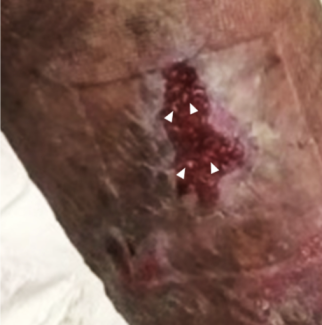
A 24-year-old male presented to the emergency department with an open, right index finger injury that occurred while playing basketball (Figure 1). Sensation and perfusion to the patient’s hand were noted to be intact. Bedside reduction under digital block was attempted without success. Given the irreducible nature of his dorsal proximal interphalangeal joint (PIPJ) dislocation, the patient was taken to the operating room for washout and reduction. A volar approach to the joint confirmed a dorsal PIPJ dislocation with disruption of the volar plate and collateral ligaments. The flexor digitorum superficialis (FDS) and flexor digitorum profundus (FDP) tendons were displaced to the radial side of the proximal phalangeal head (Figure 2). These could be seen acting as a noose on the neck of the proximal phalanx with attempted reduction. The index finger was flexed to reduce tension on the FDS and FDP tendons. The FDS was repositioned volar to the phalangeal head followed by the FDP. Gentle traction was then placed on the finger with subsequent reduction of PIPJ. Passive range of motion demonstrated a stable reduction with supporting evidence of static and live orthogonal fluoroscopic views (Figure 3). The patient was started on immediate active motion postoperatively with buddy taping of the index to the middle finger. Active motion was as tolerated with the guidance of a certified hand therapist once per week. At 3 months postoperatively, he had full flexion of the finger with a 10-degree contracture at the PIP joint. He returned to work and recreational activities with no limitations.


fluoroscopic views.
Q1. What are the recognized patterns of PIPJ dislocation, and which structures are characteristically damaged?
PIPJ dislocations can occur in the dorsal, volar, or lateral plane. These are common injuries in the setting of contact sports.1 Each type of PIPJ dislocation is named according to the distal fragment relative to the PIP joint. A volar PIPJ dislocation can be recognized as either simple or rotatory. A simple volar dislocation is caused by rupture of the central slip. A rotatory volar dislocation is a more complex injury, involving rupture of one of the collateral ligaments and entrapment of the proximal phalanx condyle between the central slip and lateral band. Dorsal dislocations are considered the most common of PIPJ dislocations. The combination of joint hyperextension with longitudinal compression results in tearing of the collateral ligaments and volar plate avulsion off the middle phalanx with or without associated fracture. Dorsal dislocations are further classified into simple or complex based on whether there is contact between the middle phalanx and the condyles of the proximal phalanx. Lateral dislocations are caused by disruption of the collateral ligament with possible volar plate avulsion.2,3
Q2. What are the causes of irreducible PIPJ dislocation?
Irreducible PIPJ dislocations can present in any plane of injury. The cause of failed closed reduction is typically disruption and interposition of nearby supporting structures. In the rotatory volar dislocation, this is usually due to unicondylar proximal phalanx entrapment between the central slip and volarly displaced lateral bands. In the dorsal plane, irreducible dislocations are commonly caused by the ruptured volar plate interposed within the joint or displacement of the flexor tendons. In the lateral plane, irreducible dislocations are caused by interposition of the lateral band.4,5
Q3. What is the course of management and treatment for a complex dorsal PIPJ dislocation?
Irreducible complex dorsal dislocations are treated through open reduction. This allows proper visualization of all structures, adequate repair, and realignment of the disrupted joint space.6 The open reduction technique is usually approached from a dorsal incision between the lateral band and central slip. A Freer elevator is then utilized to relocate the volar plate from between the joint, thus allowing realignment into proper position. Joint stability post reduction is assessed with physical exam and imaging. If the joint remains stable with full range of motion, movement of the joint is encouraged with supportive buddy taping. If the joint is successfully reduced but remains unstable, extension block splinting is recommended. The joint is placed in a splint that prevents extension past the point where the joint may dislocate. The joint is progressively extended by 10 degrees weekly for 3 weeks or until the joint becomes stable.5 In our case, the patient presented with a volar wound, so this was used to access the PIPJ. Using this incision, we were able to adequately visualize the radially displaced tendons acting like a noose on the head of the proximal phalanx. Reduction was performed by repositioning the tendons over the phalanges in the midline. Following this, gentle traction allowed stable reduction of the joint.
Q4. What are common complications following treatment of complex PIPJ dislocation?
Complications can arise following both open and closed reductions. These include joint stiffness, chronic arthritis, septic arthritis, and swan neck or boutonniere deformity.7-9 Multiple attempts at closed reductions or repetitive injury following successful closed reduction can lead to joint stiffness and chronic arthritis. If open reduction is required, then proper sterile technique, debridement, and antibiotics as indicated are important in preventing joint infection and possible septic arthritis.8 If appropriate splinting and therapy protocol are not followed, joint deformity can occur. A swan neck deformity, usually seen in dorsal dislocations, is recognized by a PIPJ hyperextension and distal interphalangeal joint (DIPJ) flexion.In contrast, a boutonniere deformity, usually seen in volar dislocations, presents with PIPJ hyperflexion and DIPJ hyperextension.9
In summary, dorsal PIPJ dislocations are common sports injuries. In an irreducible, dorsal dislocation, the degree of displacement causing loss of condylar contact and intervening injured structures limits closed reduction attempts. Open reduction allows for adequate visualization of the affected joint; anatomic re-alignment of tendons; and repair as indicated for volar plate, collateral ligaments, and the neurovascular bundle. Of critical importance is the postoperative therapy protocol, emphasizing early motion to prevent adhesions and joint stiffness.
Acknowledgments
Affiliations: 1Michigan State University, College of Human Medicine, Grand Rapids, Michigan; 2Integrated Plastic Surgery Residency, Corewell Health/Michigan State University, Grand Rapids, Michigan; 3Elite Hand and Plastic Surgery Center, Grand Rapids, Michigan
Correspondence: Olivia Means, MD; omeans7@gmail.com
Disclosure: The authors disclose no financial or other conflicts of interest.
References
- Borchers JR, Best TM. Common finger fractures and dislocations. Am Fam Physician. 2012;85(8):805-810.
- Elfar J, Mann T. Fracture-dislocations of the proximal interphalangeal joint. J Am Acad Orthop Surg. 2013;21(2):88-98. doi:10.5435/JAAOS-21-02-88
- Taqi M, Collins A. Finger Dislocation. [Updated 2022 Nov 20]. In: StatPearls [Internet]. Treasure Island (FL): StatPearls Publishing; 2023 Jan-. Available from: https://www.ncbi.nlm.nih.gov/books/NBK551508/
- Saitta BH, Wolf JM. Treating proximal interphalangeal joint dislocations. Hand Clin. 2018;34(2):139-148. doi:10.1016/j.hcl.2017.12.004
- Eaton RG, Littler JW. Joint injuries and their sequelae. Clin Plast Surg. 1976;3(1):85-98.
- Wolfe SW, Pederson WC, Kozin SH, Cohen MS. Dislocations and Ligament Injuries of the Digits. In: Green's Operative Hand Surgery. 8th ed. Elsevier, Philadelphia, PA; 2017: 326-364.
- Stern PJ, Lee AF. Open dorsal dislocations of the proximal interphalangeal joint. J Hand Surg. 1985;10(3): 364-70. doi:10.1016/s0363-5023(85)80036-8
- Lane R, Nallamothu SV. Swan-Neck Deformity. In: StatPearls. Treasure Island (FL): StatPearls Publishing; June 26, 2023.
- Fox PM, Chang J. Treating the proximal interphalangeal joint in swan neck and boutonniere deformities. Hand Clin. 2018;34(2):167-176. doi:10.1016/j.hcl.2017.12.006


