Tip 1: Take Better Office Photo
Utilizing a few tips can help you take markedly improved clinical patient photographs. First, back up (physically, not technically).
The benefits of backing away from the subject include:
1. To avoid “camel face” or “fish-eye” effect, back away from the subject and either:
• Zoom in to take the photo, or take the photo from a distance and crop the final image on the computer.
2. To avoid overexposure when using a flash, back up, take the photo and crop the final image.
• If you are planning to crop, use an image size of at least 3 megapixels to avoid loss of detail when later enlarged.
Quick color correction: To easily correct color in a photo when there is a an abnormal hue (red, yellow, blue, green) to the entire image:
• Place a white ruler (or other object) in the field before taking the photo.
• On the computer in editing mode, use the color cast (also called white balance) editing tool.
Multiple shots: Taking more than one shot is the key to a well-focused image
• Take at least 2 photos every time to ensure at least one with good focus. When you review on your computer, delete all but the best. This is especially important when photographing children.
Keep your elbows in: To hold the camera steady and reduce blur, press your elbows into your side to steady your arms while shooting.
Tangential lighting: To show elevation and enhance surface detail, use tangential lighting from a wall lamp or window and avoid using a flash.
Simple, inexpensive dermoscopic photos: To take photos through the dermoscope (and microscope) using a digital camera:
• Swab the lesion with alcohol to eliminate surface scale reflection.
• On camera, use macro setting and no flash.
• Use the focus spacer of dermoscope – place it lightly on the lesion and avoid pressure on the lesion especially if you are trying to capture the vessels.
• Place the camera gently against dermoscope, checking image on the screen, and shoot. You can zoom with the camera if desired, or on the computer.
Elizabeth O’Brien, MD
Montreal, Quebec, Canada
 Tip 2: Delivering TCA for Scars & Pores
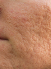
For dilated pores (eg, dilated pore of Winer) or ice-pick acne scarring, we often use 80% to 90% Trichloroacetic acid (TCA) applied into the narrow opening of the scar or pore. I now find that dunking a sharp hyfrecator needle into the TCA is a great delivery vehicle for depositing TCA into the treated lesion.
Benjamin Barankin, MD
Toronto, ON, Canada
Tip 3: “Seen many Derms”
It seems like Lichen simplex chronicus is not something a dermatologist would have trouble recognizing, nor would it be something for which many dermatologists would prescribe the wrong treatment. “Seen many Derms” makes me wonder about very poor compliance. If that is an issue, intralesional Kenalog or daily “phototherapy” visits at which clobetasol is also applied could be useful strategies.
Steve Feldman, MD, PhD
Winston-Salem, NC
 Tip 4: Welcome-to-the-Practice Pack
A welcoming pack is a good gesture toward new persons joining your practice. In healthcare centers, whether hospitals or clinics, there is a flow of personnel, and always there is a flow of newcomers. Those joining the organization recently need a good orientation to the place, especially if it is large. Organizing a comprehensive folder of documents on necessary information about the organization is very important to speed up the orientation process and also to give a good impression about the place. This pack may contain the phone directory, the organizational chart, important safety measures, general policy and procedure of the clinic, etc.
Khalid Al Aboud, MD
Makkah, Saudi Arabia
 Tip 5: Aftercare Access
For patients who have procedures done at the office, it can be a nice policy to offer them your email to contact you if they have any questions or concerns. Patients are quite pleased, and less likely to report problems with the procedure.
Benjamin Barankin, MD
Toronto, Ontario, Canada
Dr. Barankin is a dermatologist based in Toronto, Canada. He is author-editor of six books in dermatology and is widely published in the dermatology and humanities literature.
He is also co-editor of Dermanities (dermanities.com), an online journal devoted to the humanities as they relate to dermatology.
Tip 1: Take Better Office Photo
Utilizing a few tips can help you take markedly improved clinical patient photographs. First, back up (physically, not technically).
The benefits of backing away from the subject include:
1. To avoid “camel face” or “fish-eye” effect, back away from the subject and either:
• Zoom in to take the photo, or take the photo from a distance and crop the final image on the computer.
2. To avoid overexposure when using a flash, back up, take the photo and crop the final image.
• If you are planning to crop, use an image size of at least 3 megapixels to avoid loss of detail when later enlarged.
Quick color correction: To easily correct color in a photo when there is a an abnormal hue (red, yellow, blue, green) to the entire image:
• Place a white ruler (or other object) in the field before taking the photo.
• On the computer in editing mode, use the color cast (also called white balance) editing tool.
Multiple shots: Taking more than one shot is the key to a well-focused image
• Take at least 2 photos every time to ensure at least one with good focus. When you review on your computer, delete all but the best. This is especially important when photographing children.
Keep your elbows in: To hold the camera steady and reduce blur, press your elbows into your side to steady your arms while shooting.
Tangential lighting: To show elevation and enhance surface detail, use tangential lighting from a wall lamp or window and avoid using a flash.
Simple, inexpensive dermoscopic photos: To take photos through the dermoscope (and microscope) using a digital camera:
• Swab the lesion with alcohol to eliminate surface scale reflection.
• On camera, use macro setting and no flash.
• Use the focus spacer of dermoscope – place it lightly on the lesion and avoid pressure on the lesion especially if you are trying to capture the vessels.
• Place the camera gently against dermoscope, checking image on the screen, and shoot. You can zoom with the camera if desired, or on the computer.
Elizabeth O’Brien, MD
Montreal, Quebec, Canada
 Tip 2: Delivering TCA for Scars & Pores
For dilated pores (eg, dilated pore of Winer) or ice-pick acne scarring, we often use 80% to 90% Trichloroacetic acid (TCA) applied into the narrow opening of the scar or pore. I now find that dunking a sharp hyfrecator needle into the TCA is a great delivery vehicle for depositing TCA into the treated lesion.
Benjamin Barankin, MD
Toronto, ON, Canada
Tip 3: “Seen many Derms”
It seems like Lichen simplex chronicus is not something a dermatologist would have trouble recognizing, nor would it be something for which many dermatologists would prescribe the wrong treatment. “Seen many Derms” makes me wonder about very poor compliance. If that is an issue, intralesional Kenalog or daily “phototherapy” visits at which clobetasol is also applied could be useful strategies.
Steve Feldman, MD, PhD
Winston-Salem, NC
 Tip 4: Welcome-to-the-Practice Pack
A welcoming pack is a good gesture toward new persons joining your practice. In healthcare centers, whether hospitals or clinics, there is a flow of personnel, and always there is a flow of newcomers. Those joining the organization recently need a good orientation to the place, especially if it is large. Organizing a comprehensive folder of documents on necessary information about the organization is very important to speed up the orientation process and also to give a good impression about the place. This pack may contain the phone directory, the organizational chart, important safety measures, general policy and procedure of the clinic, etc.
Khalid Al Aboud, MD
Makkah, Saudi Arabia
 Tip 5: Aftercare Access
For patients who have procedures done at the office, it can be a nice policy to offer them your email to contact you if they have any questions or concerns. Patients are quite pleased, and less likely to report problems with the procedure.
Benjamin Barankin, MD
Toronto, Ontario, Canada
Dr. Barankin is a dermatologist based in Toronto, Canada. He is author-editor of six books in dermatology and is widely published in the dermatology and humanities literature.
He is also co-editor of Dermanities (dermanities.com), an online journal devoted to the humanities as they relate to dermatology.
Tip 1: Take Better Office Photo
Utilizing a few tips can help you take markedly improved clinical patient photographs. First, back up (physically, not technically).
The benefits of backing away from the subject include:
1. To avoid “camel face” or “fish-eye” effect, back away from the subject and either:
• Zoom in to take the photo, or take the photo from a distance and crop the final image on the computer.
2. To avoid overexposure when using a flash, back up, take the photo and crop the final image.
• If you are planning to crop, use an image size of at least 3 megapixels to avoid loss of detail when later enlarged.
Quick color correction: To easily correct color in a photo when there is a an abnormal hue (red, yellow, blue, green) to the entire image:
• Place a white ruler (or other object) in the field before taking the photo.
• On the computer in editing mode, use the color cast (also called white balance) editing tool.
Multiple shots: Taking more than one shot is the key to a well-focused image
• Take at least 2 photos every time to ensure at least one with good focus. When you review on your computer, delete all but the best. This is especially important when photographing children.
Keep your elbows in: To hold the camera steady and reduce blur, press your elbows into your side to steady your arms while shooting.
Tangential lighting: To show elevation and enhance surface detail, use tangential lighting from a wall lamp or window and avoid using a flash.
Simple, inexpensive dermoscopic photos: To take photos through the dermoscope (and microscope) using a digital camera:
• Swab the lesion with alcohol to eliminate surface scale reflection.
• On camera, use macro setting and no flash.
• Use the focus spacer of dermoscope – place it lightly on the lesion and avoid pressure on the lesion especially if you are trying to capture the vessels.
• Place the camera gently against dermoscope, checking image on the screen, and shoot. You can zoom with the camera if desired, or on the computer.
Elizabeth O’Brien, MD
Montreal, Quebec, Canada
 Tip 2: Delivering TCA for Scars & Pores
For dilated pores (eg, dilated pore of Winer) or ice-pick acne scarring, we often use 80% to 90% Trichloroacetic acid (TCA) applied into the narrow opening of the scar or pore. I now find that dunking a sharp hyfrecator needle into the TCA is a great delivery vehicle for depositing TCA into the treated lesion.
Benjamin Barankin, MD
Toronto, ON, Canada
Tip 3: “Seen many Derms”
It seems like Lichen simplex chronicus is not something a dermatologist would have trouble recognizing, nor would it be something for which many dermatologists would prescribe the wrong treatment. “Seen many Derms” makes me wonder about very poor compliance. If that is an issue, intralesional Kenalog or daily “phototherapy” visits at which clobetasol is also applied could be useful strategies.
Steve Feldman, MD, PhD
Winston-Salem, NC
 Tip 4: Welcome-to-the-Practice Pack
A welcoming pack is a good gesture toward new persons joining your practice. In healthcare centers, whether hospitals or clinics, there is a flow of personnel, and always there is a flow of newcomers. Those joining the organization recently need a good orientation to the place, especially if it is large. Organizing a comprehensive folder of documents on necessary information about the organization is very important to speed up the orientation process and also to give a good impression about the place. This pack may contain the phone directory, the organizational chart, important safety measures, general policy and procedure of the clinic, etc.
Khalid Al Aboud, MD
Makkah, Saudi Arabia
 Tip 5: Aftercare Access
For patients who have procedures done at the office, it can be a nice policy to offer them your email to contact you if they have any questions or concerns. Patients are quite pleased, and less likely to report problems with the procedure.
Benjamin Barankin, MD
Toronto, Ontario, Canada
Dr. Barankin is a dermatologist based in Toronto, Canada. He is author-editor of six books in dermatology and is widely published in the dermatology and humanities literature.
He is also co-editor of Dermanities (dermanities.com), an online journal devoted to the humanities as they relate to dermatology.
 Tip 2: Delivering TCA for Scars & Pores
 Tip 4: Welcome-to-the-Practice Pack
 Tip 5: Aftercare Access


 Tip 2: Delivering TCA for Scars & Pores
 Tip 4: Welcome-to-the-Practice Pack
 Tip 5: Aftercare Access


