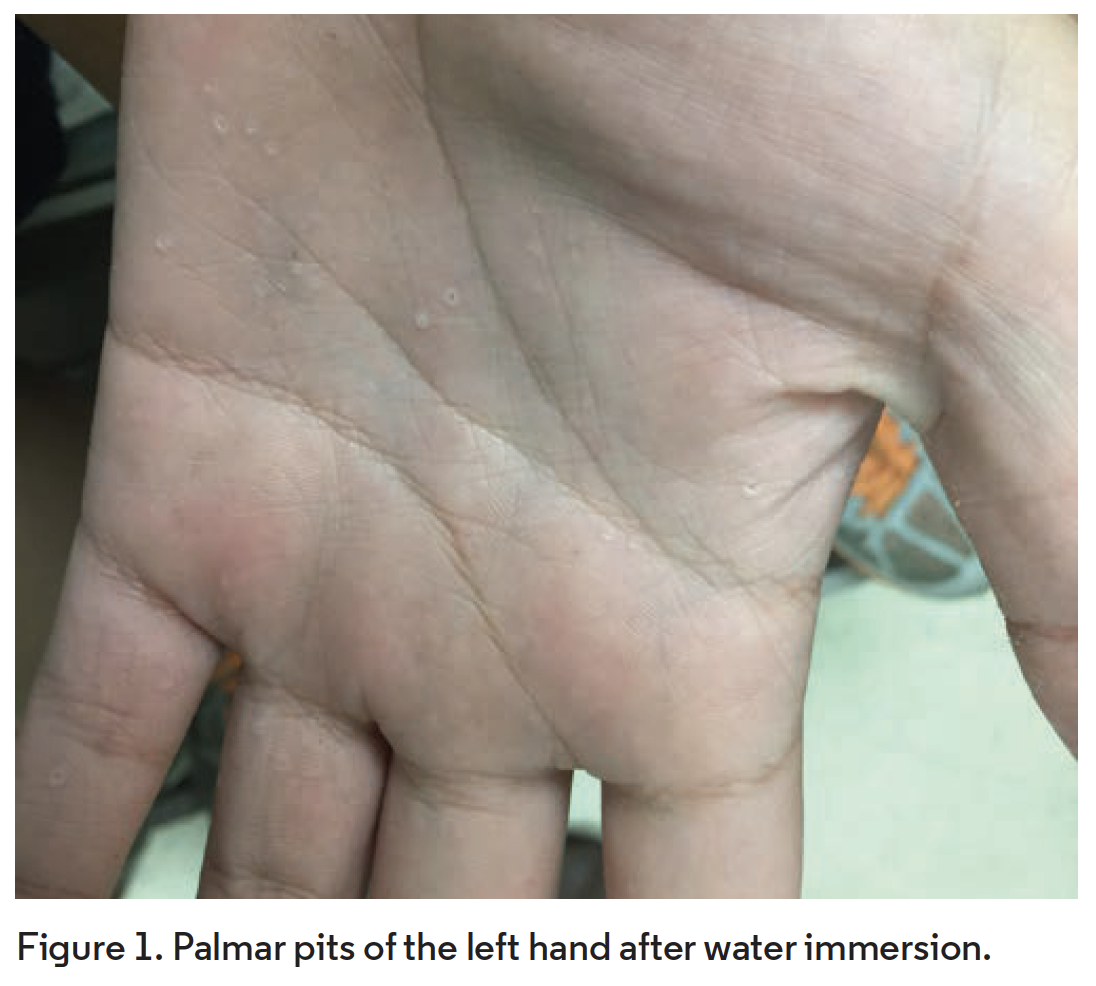
What Syndrome Caused These Palmar Pits?
Case Report
A 14-year-old Hispanic boy presented to the dermatology clinic with multiple asymptomatic, circular indentations on both palms after immersing his hands in water (Figures 1 and 2). Also present on examination was a skin-colored nodule on his left lower lip (Figure 3) and macrocephaly. A recent computed tomography (CT) maxillofacial scan performed for dental reasons demonstrated multiple maxillary and mandibular cysts in addition to calcifications of the falx cerebri and tentorium (Figure 4). Ventriculomegaly was also identified. His family history included two sisters with similar findings, an unaffected mother, and an unknown medical history for the father.

Diagnosis
Nevoid basal cell carcinoma syndrome (Gorlin syndrome)
Nevoid basal cell carcinoma syndrome (NBCCS), also known as Gorlin syndrome, is a rare genetic syndrome with a prevalence between 1 in 31,000 and 1 in 164,000.1,2 It was first described in 1894 and was further characterized by Robert Gorlin and Robert Goltz in 1960. Most cases are due to an inherited autosomal dominant loss-of-function mutation in the PTCH1 gene on chromosome 9p22.3 Mutations in SUFU and PTCH2 can also cause similar phenotypes.3 The syndrome has variable expressivity and near complete penetrance.3,4
NBCCS can affect many organ systems but particularly predisposes the patient to develop multiple basal cell carcinomas (BCCs) at an early age. There is a racial predilection for BCC development, with African American patients with NBCCS presenting with fewer BCCs than White patients. The incidence of BCCs in patients with NBCCS appears to be equal between affected men and women.5
Clinical Presentation
There are more than 100 recognized features of NBCCS. The major clinical features include the early development of multiple BCCs, jaw odontogenic keratocysts (OKCs), palmar and planar pits, lamellar calcification of the falx cerebri, and a family history of first-degree relatives with NBCCS (Figures 1-4). These classic symptoms usually present in the late teens or early adulthood.3

Multiple BCCs and palmar/planter pits are the dermatologic hallmarks of the disease. The number of BCCs can vary from a few to thousands. One study found that 80% of affected White patients had at least one BCC, with their first tumor appearing at the average age of 23 years, while 38% of affected African American patients had at least one BCC with an average age of first appearance at 21 years old.6 BCCs in NBCCS are most commonly the nodular subtype and do not differ histologically from sporadic BCCs.7 Palmar and planter pits are highly characteristic of NBCC and are present in up to 80% of patients.2 These pits are asymptomatic, shallow well-demarcated depressions in the skin. They are flesh or red-colored, permanent
lesions that do not wax and wane. They may become more pronounced after soaking the hands and feet in water.2

OKCs, cystic bone lesions lined by keratinized epithelium, are present in approximately 90% of individuals with PTCH1-related NBCCS.3 These cysts are clinically aggressive with a high reoccurrence rate.6 OKCs in NBCCS are often multiple and peak in the second and third decade.2 They may be incidentally detected on routine dental exam or may present with jaw swelling, mild pain, or altered taste.8 Other characteristic findings include calcification of the falx cerebri, which is seen in about 65% of affected patients and 80% to 90% of affected patients over the age of 40.6 Calcifications can be identified with a skull x-ray.

Minor features of the disease include characteristic facies, brain tumors, myogenic tumors, skeletal abnormalities, cardiac or ovarian fibromas, lymphomesenteric cysts, and ocular anomalies. The characteristic facies consist of frontal bossing, macrocephaly, hypertelorism, high-arched eyebrows and palate, widened nasal bridge, and mandibular prognathism.8 Macrocephaly is often the first feature to be observed in a large proportion of babies.3 Early disease recognition in children is particularly important due to increased risk of childhood brain tumors. Medulloblastomas develop in 3% to 5% of patients with NBCCS and may present in a child at a younger age than usual (younger than 5 years).9 Skeletal abnormalities are commonly seen in NBCCS and include bifid ribs, marked widening or the anterior rib ends, and fusion and modelling defects of the ribs.6

Differential Diagnosis
Palmer and planter pits may be seen in common conditions such as punctate keratoderma, pitted keratolysis, and psoriasis. Less common causes of palmer pits include Darier disease, an autosomal dominant syndrome which presents with wart-like lesions and mucosal changes.10 Multiple BCCs have been described in other rare genetic syndromes, such as Bazex syndrome and Rombo syndrome.3 However, palmar pits in the presence of multiple BCCs in a younger individual is highly suggestive of NBCCS. Clinical suspicion should be followed by radiologic studies to look for skeletal abnormalities. The diagnosis of NBCCS is made clinically based on fulfillment of two major diagnostic criteria or one major and two minor diagnostic criteria (Table). Genetic testing may be useful when the diagnosis cannot be established clinically.8

Management and Treatment
Diagnostic workup includes baseline measurement of head circumference, physical examination for birth defects, x-rays to evaluate for rib/vertebral anomalies and falx calcification, ophthalmologic evaluation, dental evaluation, and skin evaluation.3 Following diagnosis, close surveillance and regular follow up is recommended for detection of additional tumors. Genetic counseling may be valuable for patients and their families.
Treatment options for BCCs in NBCCS are the same as for sporadic BCCs. Surgical excision, specifically Mohs micrographic surgery, remains the first-line therapy. Curettage and electrodesiccation, laser therapy, topical therapies, and photodynamic therapy have also been tried.8 Treatment with sonic hedgehog inhibitor vismodegib has been shown to reduce tumor burden and block growth of new BCCs compared with placebo in patients with NBCCS.11 However, its side effects may be prohibitive in its widespread use. Of note, radiation therapy in patients with NBCCS is not recommended, as studies have shown that the use of radiotherapy can induce BCC development.12,13 Additionally, patients should be advised to limit sun exposure as UV light may promote tumorigenesis in NBCCS.14
Our Patient
Genetic analysis was performed and confirmed a PTCH1 mutation. His family history consisted of two sisters with similar findings. Their mother did not demonstrate the phenotype and the medical history of the father was unknown. It is likely that the father carried the mutation, as 70% to 80% of patients diagnosed with NBCCS have an affected parent.3 Our patient fulfilled four of the six major diagnostic criteria of NBCCS. He was counseled to avoid sun exposure and to annually visit the dermatologist for a skin cancer screening.
Conclusion
Our case presents a unique opportunity to review an uncommon genetic syndrome that may present as asymptomatic palmar pits in clinic. NBCCS is a hereditable autosomal dominant condition that includes early development of multiple BCCs, jaw OKCs, palmar and plantar pits, lamellar calcification of the falx cerebri, and a positive family history. Clinicians should be aware of the classical vignette of NBCCS, as prompt diagnosis may lead to early treatment of neoplasms and reduction of long-term sequelae.
References
1. Evans D, Howard E, Giblin C, et al. Birth incidence and prevalence of tumor-prone syndromes: estimates from a UK family genetic register service. Am J Med Genet A. 2010;152A(2):327-332. doi:10.1002/ajmg.a.33139
2. Shanley S, Ratcliffe J, Hockey A, et al. Nevoid basal cell carcinoma syndrome: review of 118 affected individuals. Am J M Genet. 1994;50(3):282-290. doi:10.1002/ajmg.1320500312
3. Evans DG, Farndon, PA. Nevoid basal cell carcinoma syndrome. In: Adam MP, Ardinger HH, Pagon RA, et al. GeneReviews. Updated March 29, 2018. Accessed August 31, 2021. http://ncbi.nlm.nih.gov/books/NBK1151
4. Anderson DE, Taylor WB, Falls HF, Davidson RT. The nevoid basal cell carcinoma syndrome. Am J Hum Genet. 1967;19(1):12-22.
5. Jones EA, Sajid MI, Shenton A, Evans DG. Basal cell carcinomas in Gorlin syndrome: a review of 202 patients. J Skin Cancer. 2011;2011:217378. doi:10.1155/2011/217378
6. Kimonis VE, Goldstein AM, Pastakia B, et al. Clinical manifestations in 105 persons with nevoid basal cell carcinoma syndrome. Am J Med Genet. 1997;69(3):299-308. doi:10.1002/(sici)1096-8628(19970331)69:33.0.co;2-m
7. Rehefeldt-Erne S, Nägeli MC, Winterton N, et al. Nevoid basal cell carcinoma syndrome: report from the Zurich nevoid basal cell carcinoma syndrome cohort. Dermatology. 2016;232(3):285-292. doi:10.1159/000444792
8. Barankin B, Goldenberg G. Nevoid basal cell carcinoma syndrome (Gorlin syndrome). UpToDate. Updated April 16, 2021. Accessed August 31, 2021. https://uptodate.com/contents/nevoid-basal-cell-carcinoma-syndrome-gorlin-syndrome?search=nevus%20basal%20cell%20carcinoma&source=search_result&selectedTitle=1~41&usage_type=default&display_rank=1#H9
9. Evans DG, Farndon PA, Burnell LD, Gattamaneni HR, Birch JM. The incidence of Gorlin syndrome in 173 consecutive cases of medulloblastoma. Br J Cancer. 1991;64(5):959-961. doi:10.1038/bjc.1991.435
10. Suryawanshi H, Dhobley A, Sharma A, Kumar P. Darier disease: a rare genodermatosis. J Oral Maxillofac Pathol. 2017;21(2):321. doi:10.4103/jomfp.jomfp_170_16
11. Tang JY, Mackay-Wiggan JM, Aszterbaum M, et al. Inhibiting the hedgehog pathway in patients with the basal-cell nevus syndrome. N Engl J Med. 2012;366(23):2180-2188. doi:10.1056/nejmoa1113538
12. Stavrou T, Bromley CM, Nicholson HS, et al. Prognostic factors and secondary malignancies in childhood medulloblastoma. J Pediatr Hematol Oncol. 2001;23(7):431-436. doi:10.1097/00043426-200110000-00008
13. O’Malley S, Weitman D, Olding M, Sekhar L. Multiple neoplasms following craniospinal irradiation for medulloblastoma in a patient with nevoid basal cell carcinoma syndrome. J Neurosurg. 1997;86(2):286-288. doi:10.3171/jns.1997.86.2.0286
14. Chiang A, Jaju PD, Batra P, et al. Genomic stability in syndromic basal cell carcinoma. J Invest Dermatol. 2018;138(5):1044-1051. doi:10.1016/j.jid.2017.09.048


