What Is the Cause of These Numerous Papules and Nodules on the Skin?
Case Report

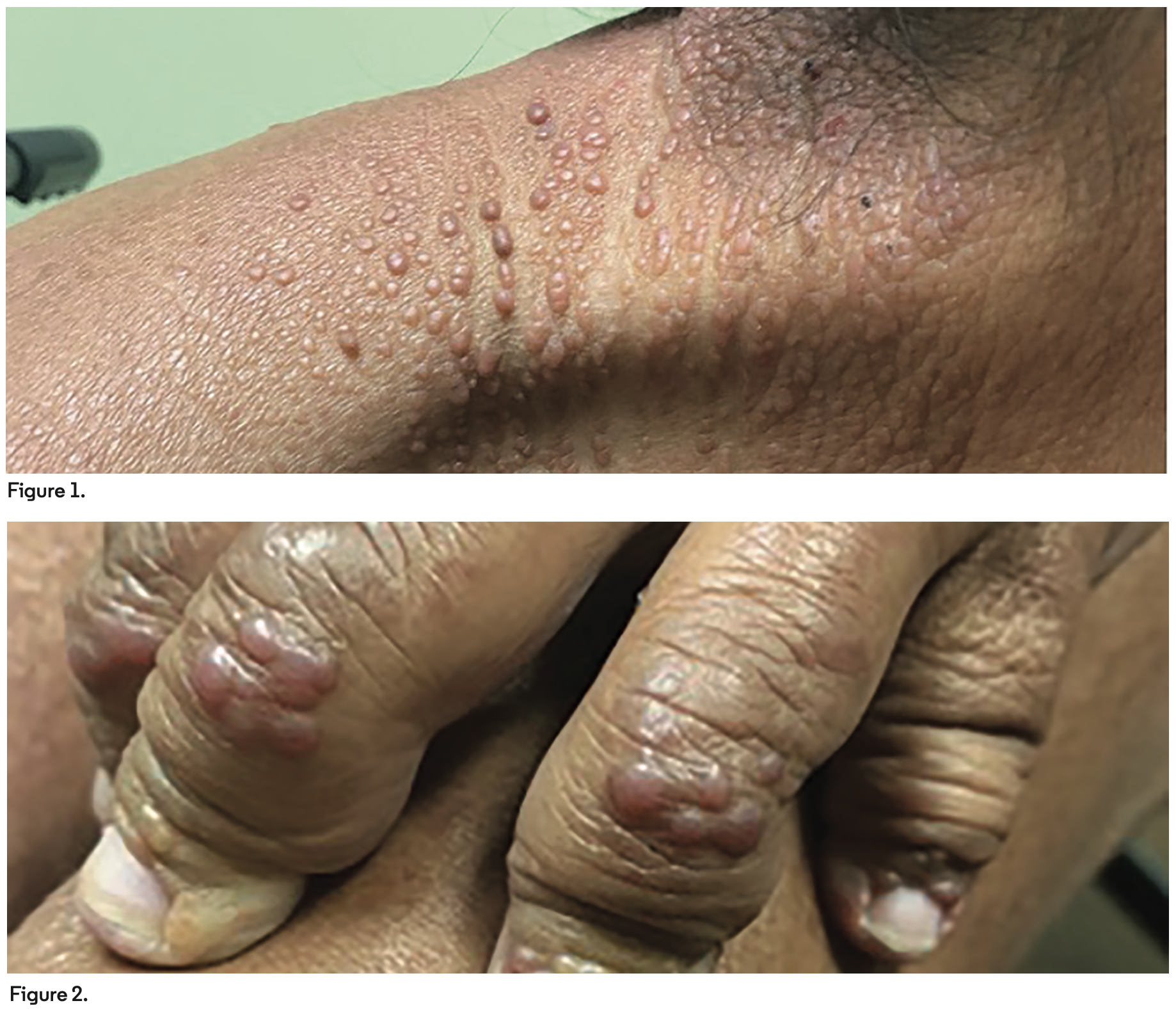
A 50-year-old woman presented with a progressive and generalized papulonodular eruption associated with debilitating arthralgias. Many smooth, coral-red, beadlike papules and nodules were arrayed linearly on the neck and extremities, as well as the nail folds and metacarpophalangeal joints (Figures 1 and 2). Over 6 years, she had developed numerous phalangeal fractures resulting in severe hand deformities.
What Is The Diagnosis?
Keep scrolling for the answer!
Diagnosis:
 Multicentric Reticulohistiocytosis
Multicentric reticulohistiocytosis (MRH) is a rare inflammatory disease with unknown etiology that typically affects White women in the fourth decade of life.1 It affects women at a rate of 3:1 compared with men.1 At this time, there is no published data on the prevalence and incidence of MRH, and it has largely been reported in case studies.2 It is classified as a non-Langerhans cell histiocytosis.3 MRH is characterized by its musculoskeletal and cutaneous features. Musculoskeletal manifestations occur as a symmetric and erosive severe arthropathy that affects the distal interphalangeal, tibiofemoral, and glenohumeral joints, feet, and elbows. Left untreated or poorly controlled, it can progress to arthritis mutilans. Cutaneous features consist of erosive nodules and multiple papules. Coral beading is a pathognomonic feature of the disease demonstrated by small periungual papules in a linear array.4
Clinical Presentation and Histology
Most commonly, arthralgia in the form of an aggressive erosive peripheral polyarthritis typically precedes cutaneous findings by 3 years.1 Arthritis mutilans is present on imaging in more than half of cases, and the features can range from self-limiting to relapsing-remitting.1 Cutaneous manifestations appear as reddish-brown to flesh-colored papules and nodules, ranging in size from 1 mm to 1 cm or greater. Some are confluent and form a cobblestone appearance. The most commonly affected areas are the face and dorsal hands.1 In rarer cases, cardiopulmonary involvement may occur and result in pleural and pericardial effusions.5-9 Even more uncommon is liver involvement.10
The histologic hallmark of advanced dermal lesions is multinucleated giant cells with a ground-glass eosinophilic cytoplasm.4,11 MRH histiocytes stain positive with periodic acid Schiff, Vimentin, CD68, CD45, MAC387, and human alveolar macrophage 56. Importantly, S100 protein, CD1a, and factor XIIIa staining are negative.4
There is no definitive laboratory test for MRH. However, certain tests are useful for ruling out other diseases with overlapping features, such as rheumatoid arthritis (ie, rheumatoid factor and anticitrullinated peptide antibodies), systemic lupus erythematosus (ie, antinuclear antibody, C3, C4), Sjögren syndrome (ie, anti-Ro and anti-La antibody), inflammatory myositis (ie, creatine phosphokinase and aldolase), and gouty arthritis (ie, uric acid levels).12 Also, erythrocyte sedimentation rate, C-reactive protein level, and lipid levels have been reported to be elevated in patients with MRH.11 While none of these tests supplant the clinicopathologic diagnosis of MRH, they may be helpful for guiding treatment and defining associated features.
Disease Associations
A new diagnosis of MRH necessitates workup for an associated malignancy, autoimmune disorder, or visceral involvement. One of 4 patients with MRH will have an associated malignancy, including colon, ovarian, cervical, stomach, lung, or breast cancer.13 Notably, resolution of the malignancy does not reliably induce remission of MRH. Autoimmune disorders like Sjögren syndrome, systemic lupus erythematosus, dermatomyositis, systemic sclerosis, celiac disease, and primary biliary cirrhosis have been associated with MRH.1 As such, a thorough review of systems, with symptom-directed workup, and age-appropriate cancer screening are necessary.
Treatment
Clinical management of MRH remains challenging. The sheer paucity of MRH cases precludes large studies; therefore, well-defined treatment guidelines for this aggressive disease do not exist. First-line treatment consists of anti-inflammatory medications like nonsteroidal anti-inflammatory drugs and systemic and intra-articular corticosteroids. Limited evidence supports using disease-modifying antirheumatic drugs (DMARDs), cyclophosphamide, and cyclosporin A.1 Several case studies have demonstrated that treatment with tumor necrosis factor-α inhibitors like etanercept, adalimumab, and infliximab have improved symptoms that were refractory to DMARDs.14 Recently, bisphosphonates have been reported to improve skin lesions and arthritis symptoms, presumably through inhibition of osteoclastic activity associated with MRH.1
The disease course can be self-limiting and nondeforming, waxing and waning with spontaneous relapses and remissions, or as an irreversible, aggressive deforming and mutilating disease. Spontaneous remission often occurs within 10 years, but irreversible arthritic changes will remain.12
Our Patient
Our patient had been previously treated with multiple therapies, including topical corticosteroids, methotrexate, prednisone, etanercept, and adalimumab. All were unsuccessful at improving skin lesions or arthralgias. At presentation in our clinic, the patient reported dysphagia, weight loss, and depression/mood changes. Her most recent mammogram had been conducted 3 years prior to presentation. The patient had not had a colonoscopy, recent Papanicolaou test, or cardiac echocardiography scan. The patient denied any family history of autoimmune disease. In the setting of concerning symptoms for a potential oncologic process and the correlation between MRH and malignancy, referrals to gastroenterology and obstetrics and gynecology were made. The patient declined biopsy, resulting in a clinical diagnosis of MRH.
Conclusion
MRH is an extremely rare dermatologic and rheumatologic condition with no current consensus on incidence and prevalence of the disease. Typical presentations begin with a progressive erosive polyarthritis that occurs several years prior to cutaneous papules and nodules. Its association with malignancy should prompt age-appropriate cancer screening. It is difficult to treat and often requires multiple modalities. MRH progression remits after 8 to 10 years, but arthritic changes are irreversible.
References
1. Selmi C, Greenspan A, Huntley A, Gershwin ME. Multicentric reticulohistiocytosis: a critical review. Curr Rheumatol Rep. 2015;17(6):511. doi:10.1007/s11926-015-0511-6
2. Tariq S, Hugenberg ST, Hirano-Ali SA, Tariq H. Multicentric reticulohistiocytosis (MRH): case report with review of literature between 1991 and 2014 with in depth analysis of various treatment regimens and outcomes. Springerplus. 2016;5:180. doi:10.1186/s40064-016-1874-5
3. Zelger BW, Sidoroff A, Orchard G, Cerio R. Non-Langerhans cell histiocytoses. A new unifying concept. Am J Dermatopathol. 1996;18(5):490-504. doi:10.1097/00000372-199610000-00008
4. Tajirian AL, Malik MK, Robinson-Bostom L, Lally EV. Multicentric reticulohistiocytosis. Clin Dermatol. 2006;24(6):486-492. doi:10.1016/j.clindermatol.2006.07.010
5. Benucci M, Sulla A, Manfredi M. Cardiac engagement in multicentric reticulohistiocytosis: report of a case with fatal outcome and literature review. Intern Emerg Med. 2008;3(2):165-168. doi:10.1007/s11739-008-0102-x
6. Lambert CM, Nuki G. Multicentric reticulohistiocytosis with arthritis and cardiac infiltration: regression following treatment for underlying malignancy. Ann Rheum Dis. 1992;51(6):815-817. doi:10.1136/ard.51.6.815
7. Yee KC, Bowker CM, Tan CY, Palmer RG. Cardiac and systemic complications in multicentric reticulohistiocytosis. Clin Exp Dermatol. 1993;18(6):555-558. doi:10.1111/j.1365-2230.1993.tb01030.x
8. Bogle MA, Tschen JA, Sairam S, McNearney T, Orsak G, Knox JM. Multicentric reticulohistiocytosis with pulmonary involvement. J Am Acad Dermatol. 2003;49(6):1125-1127. doi:10.1016/s0190-9622(03)00036-7
9. Fast A. Cardiopulmonary complications in multicentric reticulohistiocytosis. Report of a case. Arch Dermatol. 1976;112(8):1139-1141.
10. Yang HJ, Ding YQ, Deng YJ. Multicentric reticulohistiocytosis with lungs and liver involved. Clin Exp Dermatol. 2009;34(2):183-185. doi:10.1111/j.1365-2230.2008.02795.x
11. Sanchez-Alvarez C, Sandhu AS, Crowson CS, et al. Multicentric reticulohistiocytosis: the Mayo Clinic experience (1980-2017). Rheumatology (Oxford). 2020;59(8):1898-1905. doi:10.1093/rheumatology/kez555
12. Toz B, Büyükbabani N, İnanç M. Multicentric reticulohistiocytosis: Rheumatology perspective. Best Pract Res Clin Rheumatol. 2016;30(2):250-260. doi:10.1016/j.berh.2016.07.002
13. El-Haddad B, Hammoud D, Shaver T, Shahouri S. Malignancy-associated multicentric reticulohistiocytosis. Rheumatol Int. 2011;31(9):1235-1238. doi:10.1007/s00296-009-1287-7
14. Zhao H, Wu C, Wu M, et al. Tumor necrosis factor antagonists in the treatment of multicentric reticulohistiocytosis: Current clinical evidence. Mol Med Rep. 2016;14(1):209-217. doi:10.3892/mmr.2016.5253


