Advertisement

Advertisement

ADVERTISEMENT

Derm Dx

What Condition Does This Woman With Scleral Dyschromia Have?

February 2021
Figure 1.
Figure 1.

Case Report
A 54-year-old woman presented for symptoms of itching. Cutaneous examination revealed urticarial dermal plaques on her neck, chest, back, arms, and legs. Incidentally, she was found to have prominent blue sclera (Figure 1). She has had five fractures during her lifetime, though none during the last several years.

What Is The Diagnosis?
Check your answer by hitting "Next" below!

,

Diagnosis: Osteogenesis Imperfecta
Osteogenesis imperfecta (OI) is a genetic disorder characterized by brittle bones, skeletal deformities, and growth retardation. OI is the most common cause of primary osteoporosis, and its incidence is between 1 in 10,000 and 1 in 20,000. It is most commonly inherited in an autosomal dominant manner; however, less common subtypes are autosomal recessive and X-linked. The manifestations of OI are often caused by defects associated with type I collagen synthesis. The diagnosis is often based on clinical features, and management is multifactorial, involving physicians and caregivers of multiple specialties.

Classification

Table 1. Classifications of OI

Sillence et al provided an initial classification of OI with four subtypes based on clinical and radiographic features (Table 1). More recently, with the ability to identify the molecular pathogenesis of OI, 15 subtypes of OI have been identified. Types I through V are predominantly autosomal dominant, whereas type VI through type XV are autosomal recessive. Rarely, other modes of inheritance, such as X-linked, have been identified in type I.

Type I tends to be mild, and type II is lethal around the time of childbirth or shortly after. Type III is the most severe nonlethal form, and type IV is of intermediate severity (Table 2). Type I and type IV, milder forms of OI, are more common in the population.
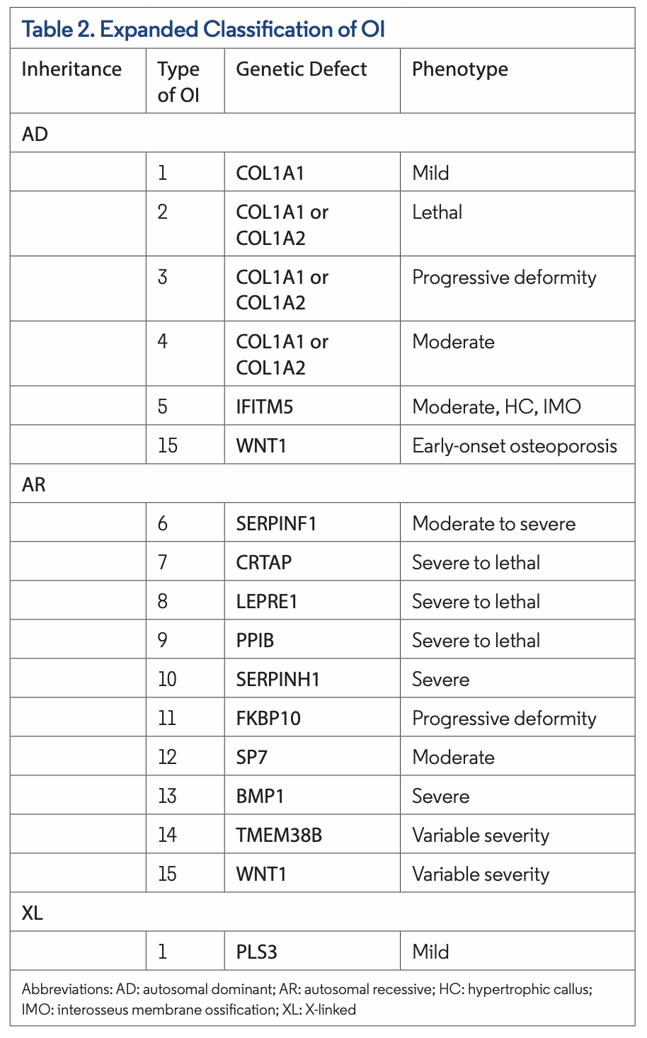
Table 2. Expanded Classification of OIClinical Presentation
The clinical presentation of OI varies based on age of onset and subtype of the disease. In the first 6 months of life, few examination findings of OI may be observed. Thereafter, the most common features are bone abnormalities, fractures, and short stature. Other common features include compression fractures, deformed craniocervical junction, hearing impairment, joint hypermobility, motor delay, osteopenia, pectus excavatum, scoliosis, teeth abnormalities (chipped teeth), and thoracic kyphosis. In addition, reduced muscle mass and strength is associated with type I, and triangular facies is associated with type III.

Similar to our patient, mucocutaneous features include blue sclera, which is often a bilateral finding (Figures 1 and 2). Blue sclerae are more common in type I OI but can also be seen in other subtypes of OI (Table 1).The blue-gray discoloration occurs due to the thin, translucent, and vascular nature of the sclera. Over time, the color of the sclera can either stay uni- form or change to a lighter blue color.

Differential Diagnosis
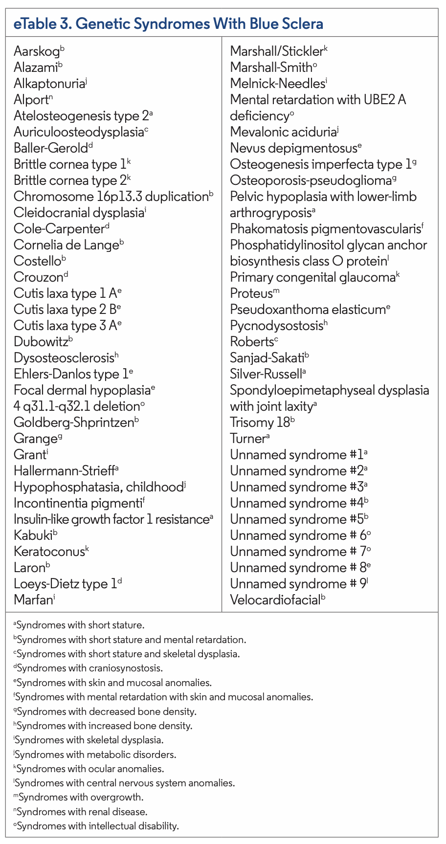
Currently, 66 genetic syndromes may present with blue sclerae and can be found in eTable 3. Nongenetic disorders and certain medications may also present with similar findings. Of the genetic syndromes, the most common to present with blue sclera include OI, Hallermann-Strieff syndrome, Kabuki syndrome, Laron syndrome, Loeys-Dietz type 1 syndrome, and Marshall-Smith syndrome. Caplan syndrome, HIV infection, hyperhomocysteinemia, iron deficiency anemia, myasthenia gravis, nevus of Ota, and acquired polyneuropathy, organo- megaly, endocrinopathy, monoclonal gammopathy and skin changes (POEMS) syndrome are nongenetic etiologies that may present with blue sclera. Additionally, minocycline, mitoxantrone, prednisone, and retigabine are medications that have been associated with blue sclera (eTable 4).

Sclera have also been observed that have shades of blue, either blue-black or blue-gray, in certain syndromes and systemic conditions. These include argyria, chrysiasis, Ehlers- Danlos syndrome Type VIIC, ochronosis, and OI. Nevus of Ota and dermal melanosis are pigmented lesions that can be blue-black or blue-gray, respectively.

,

Pathogenesis
OI has a very complicated pathogenesis that has yet to be fully elucidated. The pathogenesis in 90% of patients is secondary to an autosomal dominant structural or quantitative genetic mutation in COL1A1 or COL1A2 genes, which encode type I collagen. A defect in the synthesis of type I collagen can lead to bone deformities and systemic and oral maxillofacial malformations as observed in OI. The remaining 10% of etiologies include qualitative mutations affecting collagen processing, folding, cross-linking, posttranslational modifications, bone mineralization, and osteoblast development. Quantitative genetic mutations with normal collagen structure usually present with a milder form of the disease such as type I OI. In contrast, mutations affecting collagen structure can present with a lethal (type II), severe (type III), or moderate (type IV) forms. 

Figure 2. A closer view of the blue sclera in the right eye of the 54-year-old woman with OI.

Other Findings
Patients with OI commonly have brittle but hypodense bones. Their bone scan is typically characterized by diminished growth, hypermineralization, increased turnover, and disrupted bone architecture. Patients with OI are also at increased risk for cardiovascular disease, fractures, primary osteoporosis, and restrictive lung disease. Laboratory studies show vitamin D deficiency. Vitamin D supplementation improves symptoms. Patients who receive bisphosphonate therapy before closure of epiphyseal plates commonly present with the “zebra stripe sign,” which is associated with type I OI. Type V OI has a unique radiographic feature caused by calcified interosseous membranes and hypertrophic callus.

eTable 3. Genetic Syndromes With Blue ScleraManagement
Early diagnosis and intervention can substantially improve the clinical outcome for a patient with OI. Management of these patients is multifactorial and involves caregivers of multiple specialties, including cardiology, dentistry, genetics, orthopedics, otolaryngology, and pediatrics.

Treatment includes bisphosphonates and normalizing vitamin D levels. Bisphosphonates have been associated with increased bone mass, improved bone architecture, and decreased rate of fractures. Improved clinical outcomes are most significant among infants.

Mild OI is typically treated with the oral form of bisphosphonates while moderate to severe OI is treated intravenously. Standard guidelines for bisphosphonate dosage, frequency, and time frame of treatment remain to be established. Common side effects include acute phase reaction and hypocalcemia but are not detrimental.

Surgical repairment is indicated for patients who present with recurrent fractures or severe scoliosis.

Our Patient
Several members of our patient’s family, including her mother, grandmother, 9 of 12 siblings, and her daughter, have OI. Her daughter, who is now 22 years old, has had a total of 17 fractures, yet none in the last 4 years.

The etiology of our patient’s urticarial dermatitis could not be determined. She was treated with a 12-day tapering course of oral prednisone, topical triamcinolone 0.1% cream, and hydroxyzine (25 mg each evening). There was complete resolution of her dermatitis without recurrence.

Conclusion
OI is an inborn error of metabolism often associated with defects in type I collagen synthesis. Patients typically present with brittle bones. Blue sclera is most commonly associated with type I OI; however, it has been observed, albeit less frequently, in other types of OI. The differential diagnosis of blue sclera includes not only other genetic disorders and noninherited conditions, but it also can be an adverse effect from systemic medications. The most widely used intervention in OI is intravenous bisphosphonates. Importantly, the care of patients with OI requires the combined contributions of health care workers from various specialties. 

eTable 4. Medications Associated With Blue Sclera


,

Ms Laborada is a medical student at the University of California Riverside School of Medicine in Riverside, CA. Dr Cohen is an adjunct professor of dermatology at Touro University California College of Osteopathic Medicine in Vallejo, CA, and a dermatologist at San Diego Family Dermatology in National City, CA.

References

  1. Arundel P. Osteogenesis imperfecta. Paediatr Child Health. 2015;25(12):574-579. doi:0.1016/j.paed.2015.07.007
  2. Sillence DO, Senn A, Danks DM. Genetic heterogeneity in osteogenesis imperfecta. J Med Genet. 1979;16(2):101-116. doi:10.1136/jmg.16.2.101
  3. Valadares ER, Carneiro TB, Santos PM, Oliveira AC, Zabel B. What is new in genetics and osteogenesis imperfecta classification? J Pediatr (Rio J). 2014;90(6):536-541. doi:10.1016/j.jped.2014.05.003
  4. Tournis S, Dede AD. Osteogenesis imperfecta - a clinical update. Metabolism. 2018;80:27-37. doi:10.1016/j.metabol.2017.06.001
  5. Wilcox RA, McDonald FS. Gray-blue sclerae and osteopenia secondary to osteogenesis imperfecta. Mayo Clin Proc. 2007;82(3):265. doi:10.4065/82.3.265
  6. Brooks, JK. A review of syndromes associated with blue sclera, with inclusion of malformations of the head and neck. Oral Surg Oral Med Oral Pathol Oral Radiol. 2018;126(3):252-263. doi:10.1016/j.oooo.2018.05.012
  7. Goette A, Huth C, Behrens-Baumann W, Evert M. Severe mitral regurgitation in a patient with a bluish right sclera: an ‘extension’ of Caplan’s syndrome to the heart and eye. Eur J Intern Med. 2009;20(1):e1-e2. doi:10.1016/j.ejim.2007.06.027
  8. Marion RW, Wiznia AA, Hutcheon G, Rubinstein A. Human T-cell lymphotropic virus type III (HTLV-III) embryopathy. A new dysmorphic syndrome associated with intrauterine HTLV-III infection. Am J Dis Child. 1986;140(7):638-640. doi:10.1001/archpedi.1986.02140210036020
  9. Rathi A, Takkar B, Azad S. Ectopic lentis and blue sclera in hyperhomocysteinaemia. Natl Med J India. 2017;30(3):176.
  10. Hassen GW, Chirurgi R, Kalanrari H. Blue sclera secondary to severe iron deficiency anemia. QJM. 2017;110(12):835-836. doi:10.1093/qjmed/hcx163
  11. Wiernik PH. Blue sclerae. Lancet. 1972;2(7788):1199. doi:10.1016/s0140-6736(72)92624-4
  12. Sehgal VN, Syed NH, Aggarwal A, Sharma S, Sehgal S. Nevus of Ota/oculodermal melanocytosis: a rare report of an oral mucosal lesion involving the hard palate. Cutis. 2015;96(4):E10-E12.
  13. Limvorapitak W, Villa D. Acquired blue sclerae in POEMS syndrome. Br J Haematol. 2017;177(4):508. doi:10.1111/bjh.14646
  14. Johnston S. Feeling blue? Minocycline-induced staining of the teeth, oral mucosa, sclerae and ears – a case report. Br Dent J. 2013;215(2):71-73. doi:10.1038/sj.bdj.2013.682
  15. Reese S, Grundfast K. Minocycline-induced hyperpigmentation of tympanic membrane, sclera, teeth, and pinna. Laryngoscope. 2015;125(11):2601-2603. doi:10.1002/lary.25365
  16. Leyden MJ, Sullivan JR, Cheng ZM, Russell IS, Collins J. Unusual side effect of mitoxantrone. Med J Aust. 1983;2(10):514. doi:10.5694/j.1326-5377.1983.tb122624.x
  17. Henderson RP, Lander R. Scleral discoloration associated with long-term prednisone administration. Cutis. 1984;34(1):76-77.
  18. Clark S, Antell A, Kaufman K. New antiepileptic medication linked to blue discoloration of the skin and eyes. Ther Adv Drug Saf. 2015;6(1):15-19. doi:10.1177/2042098614560736
  19. Kim Y, Suh HS, Cha HJ, Kim SH, Jeong KS, Kim DH. A case of generalized argyria after ingestion of colloidal silver solution. Am J Ind Med. 2009;52(3):246-250. doi:10.1002/ajim.20670
  20. Smith RW, Leppard B, Barnett NL, Millward-Sadler GH, McCrae F, Cawley MI. Chrysiasis revisited: a clinical and pathologic study. Br J Dermatol. 1995;133(5):671-678. doi:10.1111/j.1365-2133.1995.tb02737.x
  21. Malfait F, De Coster P, Hausser I, et al. The natural history, including orofacial features of three patients with Ehlers-Danlos syndrome, dermatosparaxis type (EDS type VIIC). Am J Med Genet A. 2004;131(1):18-28. doi:10.1002/ajmg.a.30299
  22. Albers SE, Brozena SJ, Glass LF, Fenske NA. Alkaptonuria and ochronosis: case report and review. J Am Acad Dermatol. 1992;27(4):609-614. doi:10.1016/0190-9622(92)70230-d
  23. Azami A, Maleki N, Tavosi Z. Alkaptonuric ochronosis: a clinical study from Ardabil, Iran. Int J Rheum Dis. 2014;17(3):327-332. doi:10.1111/1756-185X.12287
  24. Shetty SR, Subhas BG, Rao KA, Castellino R. Nevus of ota with buccal mucosal pigmentation: a rare case. Dent Res J (Isfahan). 2011;8(1):52-55.
  25. Wang W, Fujiwara S, Seguchi S, Yokoyama S, Mizoguchi M. Acquired dermal melanocytosis: a case with conjunctival and gingival pigmentation. J Dermatol.  2004;31(9):752-755. doi:10.1111/j.1346-8138.2004.tb00590.x
  26. Forlino A, Marini JC. Osteogenesis imperfecta. Lancet. 2016;387(10028):1657-1671. doi:10.1016/S0140-6736(15)00728-X.

Advertisement

Advertisement

Advertisement