The Dermatopathologist

Thrombotic Retiform Purpura: A Window for Understanding Severe and Critical COVID-19 of Adults

February 2021

A 71-year-old man presented to the emergency room with significant shortness of breath. Radiographic studies demonstrated hazy bibasilar opacities that were greater in the left lung compared with the right. His low oxygenation necessitated ventilator support on March 27, 2020. Comprehensive radiographic studies of other organ systems did not disclose any abnormalities. The patient was discovered to be SARS-CoV-2 positive based on nasopharyngeal swab assessment. He was then started on hy- droxychloroquine and piperacillin/tazobactam.

The patient’s past medical history was remarkable for hypertension, ulcerative colitis that was well controlled on sulfasalazine, and gastroesophageal reflux disease.

Photomicrographs of the (A) left fingertips and (B) right toes in a patient with severe and critical COVID-19
Figure 1. Photomicrographs of the (A) left fingertips and (B) right toes in a patient with severe and critical COVID-19, who developed acral thrombosis similar to that of the patient presented, as evidenced by the digit discoloration.

His clinical course was complicated by Escherichia coli and Streptococcus bacteremia and acute kidney failure. The patient was not a candidate for other SARS-CoV-2 therapy due to the concomitant bacteremia. During his intensive care unit hospitalization, the patient developed a distinctive livedoid rash on his hands along with discoloration of his digits suggestive of acral thrombosis (Figure 1). Lesional skin was biopsied along with normal deltoid skin, the latter to assess for systemic complement activation in the event that the patient would require interventional anticomplement therapy. The patient was eventually extubated and transferred to a rehabilitation center, where he developed new symptoms of septic shock and ultimately died from complications.

Light Microscopic Findings
The biopsy of the livedoid rash from the hand demonstrated marked superficial vascular ectasia. In addition, occasional vessels including small arterioles manifested intraluminal fibrin thrombi (Figure 2). The endothelium lining the occluded blood vessels exhibited proplastic alterations. There was no frank vasculitic change as evidenced by the lack of mural fibrin deposition, angiocentric inflammation, and significant red cell extravasation.

Immunohistochemical stains were performed including a myxovirus resistant protein (MxA) stain, the surrogate marker of the type I interferon (IFN) microenvironment, which was found to be negative. Complement studies, namely C3d, C4d, and C5b-9, were conducted on the formalin-fixed paraffin-embedded tissue. The C3d stains exhibited very focal weak staining within the endothelium of
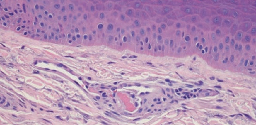
As suggested by the name, there is a pauci-inflammatory thrombogenic vasculopathy.
Figure 2. As suggested by the name, there is a pauci-inflammatory thrombogenic vasculopathy. Microvessels of the dermis exhibit bland fibrin thrombi. Significant inflammation is not seen and there- fore inflammatory vasculitic alterations are not identified (H&E 400x).

capillaries, venules, and small arterioles within the mid- and deeper dermis. The C4d pattern of endothelial cell staining showed a similar distribution to C3d but with a stronger staining intensity. The C5b-9 studies were very striking with many vessels throughout the dermis exhibiting positive staining (Figures 3 and 4). There was in excess of 45 positive blood vessels per section examined that showed an intense pattern of endothelial and subendothelial staining. Overall, the findings were those of a complement-mediated thrombotic microvascular injury syndrome. The normal deltoid skin biopsy did not show any obvious microvascular thrombosis. There were no clear-cut light microscopic abnormalities that were discernible on routine hematoxylin-eosin (H&E) stained material. Immunohistochemical stains comparable to those conducted on the livedoid skin sample were performed. The MxA stain was negative. The C3d preparation showed endothelial and subendothelial staining of four blood vessels in the mid- and deeper dermis while the C4d demonstrated six positive staining vessels in an endothelial and subendothelial array. The C5b-9 studies exhibited granular microvascular staining within the dermis with 12 positive staining blood vessels. As there were more than 10 vessels exhibiting endothelial and/or subendothelial staining for C5b-9, the deltoid biopsy established a diagnosis of systemic complement activation (Figure 4). Although the patient could be considered a candidate for eculizumab therapy given the general improvement in the patient’s clinical course, therapeutic intervention with eculizumab was not given.

In situ hybridization studies using a highly sensitive assay for SARS-CoV-2 probe were negative. In lesional and nonlesional skin, there was significant expression of SARS-CoV-2 spike glycoprotein and membrane protein within the endothelial cells lining microvessels of the dermis and subcutaneous fat (Figure 5). Additionally, there was enhanced endothelial cell expression of caspase 3 and IL-6.

The skin rash reflects systemic complement activation
Figure 3. The skin rash reflects systemic complement activation, hence the microvascular thrombosis is associated with prominent deposits of complement within the vessel walls. Illustrated is C5b-9 (diaminobenzidine [DAB] 400x).

DISCUSSION
The severe acute respiratory distress syndrome associated with coronavirus 2 (SARS-CoV-2), the etiologic agent of coronavirus disease 2019 (COVID-19), was initially identified in Wuhan, Hubei Province, China, in December 2019. It was documented to be a pandemic by the World Health Organization in early March 2020 and has since been responsible for over 75 million cases worldwide and more than 2.2 million deaths. Organ dysfunction, particularly progressive respiratory failure and a generalized coagulopathy, are associated with the highest mortality.

We demonstrated in an earlier study the important role for mannan-binding lectin pathway complement activation in the pathogenesis of severe and critical COVID-19.6 When one examines fatal cases of COVID-19 of adults, the lung samples exhibit a reproducible morphology, specifically in the context of a pauci-inflammatory necrotizing septal capillary injury syndrome. Prior studies have demonstrated conspicuous deposits of C4d and C5b-9 within the septal microvessels with evidence of colocalization of SARS- CoV-2 associated capsid proteins including spike glycoprotein and membrane and envelope capsid proteins consistent with a virally triggered complement activation syndrome as the mechanism underling COVID-19 associated microangiopathic acute respiratory distress syndrome (ARDS).

There is no evidence of a type I IFN response as revealed by the absent staining for MxA.
Figure 4. There is no evidence of a type I IFN response as revealed by the absent staining for MxA. The lack of IFN signaling despite a viral infection could contribute to the excessive viral replication in the lung (DAB, 400x).
Despite lack of any viral RNA in endothelium
Figure 5. Despite lack of any viral RNA in endothelium, there was abundant SARS-CoV-2 capsid protein including (A) spike glycoprotein (red chromagen, 1000x) and (B) capsid membrane protein (DAB, 1000x).

It has been demonstrated that in the spike glycoprotein, the critical binding site for the viral receptor, namely angiotensin converting enzyme 2 (ACE-2), has glycosylation sites for high mannose structures that is recognized by certain residues on mannan-binding lectin resulting in MASP-2 activation. The subsequent activation of MASP-2 leads to the formation of C5b-9. MASP-2 is very similar to the C1s molecule, suggesting commonality in molecular origin. The basis of the spike glycoprotein localization to capillary endothelium is attributable to ACE-2 expression in the septal microvessels of the lung. ACE-2 is also found in other microvascular beds such as the skin, explaining the localization of spike glycoprotein and components of complement activation within the skin and other organ systems as will be discussed presently.

,

Some understanding in the pathophysiology that underlies severe and critical COVID-19 can be gleaned by carefully assessing the pathologic findings in the skin in these patients. The patient presented in this anecdotal case report has a classic skin rash that is a hallmark of severe and critical COVID-19, namely COVID-19 associated thrombotic retiform purpura. COVID-19 thrombotic retiform purpura is characterized by a net-like purplish skin rash typically involving acral sites occurring almost exclusively in patients who have microangiopathic ARDS related to SARS-CoV-2.6 To date we have encountered seven cases of thrombotic retiform purpura. All patients had critical COVID-19 and were on ventilator support when they developed the acral-based livedoid rash.

The morphology and immunohistochemical profile demonstrated in this case would be considered prototypic for thrombotic retiform purpura of severe and critical COVID-19. In particular, the findings are invariably in the context of a pauci-inflammatory thrombogenic vasculopathy with involvement of capillaries, venules, and typically the arterial system. This dual venous and arterial involvement is reminiscent of antiphospholipid antibody syndrome, although these patients do not have serologic evidence of these prothrombotic antibodies. It is a complement driven process, as revealed in this case and other cases whereby there are significant microvascular granular deposits of components of complement activation including C3d, C4d, C5b-9, and the mannan-binding lectin pathway specific component of complement activation, specifically MASP-2.

The fact that there is both C4d and MASP-2 microvascular staining biopsies of COVID-19 associated thrombotic retiform purpura indicates the role for mannan-binding lectin pathway activation in the pathogenesis of severe, critical, and fatal COVID-19. A critical finding in these cases is docked viral capsid protein, including spike glycoprotein and the envelope, and membrane protein within the endothelium of the cutaneous microvessels likely as the impetus to activating the mannan-binding lectin pathway and resulting in C5b-9 mediated endothelial cell injury. In particular, the spike glycoprotein appears to travel with the capsid and membrane proteins, lodging onto ACE-2 positive endothelial cells of the cutaneous vasculature, whereby the sugar moieties of the spike glycoprotein interact with mannan-binding lectin activating the pathway leading to microvascular thrombosis. Of great interest is the fact that this spike glycoprotein endothelial cell engagement leading to mannan-binding lectin activation occurs in the context of a pseudovirion, as all in situ hybridization studies to demonstrate viral RNA have been negative. Prior studies have demonstrated that the spike glycoprotein is able to engage with ACE-2 and be endocytosed by a human cell in the absence of an intact virion. It should also be emphasized that even if there was no docking of the spike glycoprotein to ACE-2 positive microvascular beds in the skin and other organ systems such as the brain, kidney, and heart, there could still be systemic complement activation because of the massive degree of mannan-binding lectin pathway activation that occurs in the lung; activation of the mannan-binding lectin pathway will trigger the alternative pathway activation. Furthermore, complement pathway activation contributes to a significant procoagulant state because of the positive feedback between components of complement activation and the coagulation pathway.

We have also demonstrated that the microvessels of the skin and subcutaneous fat are very rich in ACE-2 expression in the endothelium likely accounting for the ability of the released spike glycoprotein and associated capsid proteins derived from intact virus from dying cells originating in the lung to bind to the endothelium of the cutaneous microvascular beds hence resulting in this distinctive skin rash in the setting of severe and critical COVID-19. Also, it should be further emphasized that ACE-2, which is the receptor for SARS-CoV-2, once engaged with spike glycoprotein either as a pseudovirions or in the context of an intact virus, is endocytosed and no longer available to perform the critical hydrolytic enzymatic conversion of angiotensin 1 into angiotensin 7, contributing to the cardiovascular demise that characterizes severe and critical CO- VID-19. Angiotensin 1 has profound vasoconstrictive properties while angiotensin 7 is associated with significant endothelial cell protective effects including vasodilation. Angiotensin 1 and angiotensin 2 have been associated with inflammation, oxidative stress, and fibrosis, and ACE-2 is involved in their deactivation. Reduced ACE-2 activity will increase angiotensin 2, leading to oxygen species formation and interference with antioxidant and vasodilatory signals such as Nox2, which further triggers complement activation.

The thrombotic retiform purpuric rashes of COVID-19 are light microscopically and mechanistically similar to other catastrophic thrombotic microvascular syndromes such as atypical hemolytic uremic syndrome and catastrophic antiphospholipid antibody syndrome.

The finding of complement deposition in the microvessels of normal deltoid skin is noteworthy and does corroborate the theory that pseudovirioncritical COVID-19 represents systemic complement activation syndromes. The deltoid skin biopsy has been utilized for a number of years at Weill Cornell Medicine to detect evidence of systemic complement activation, although in a different clinical setting, namely to rule in or out atypical hemolytic uremic syndrome. We performed the deltoid biopsy on patients with severe and critical COVID-19 to document systemic complement activation; by documenting systemic complement activation, an additional therapeutic intervention with complement inhibition therapy was possible.Invariably the deltoid skin biopsy in patients with critical COVID-19 shows significant deposits of C4d, a component of mannan-binding lectin pathway activation, and C5b-9. This finding would not be expected if the complement activation was from the excessive synthesis in the lung alone, and it supports the pseudovirion hypothesis. We have shown that deltoid skin samples from patients with severe and fatal COVID-19 exhibit docked pseudovirions, namely spike glycoprotein and other viral capsid proteins such as membrane and envelope capsid proteins within the endothelium in the absence of viral RNA albeit replicative intact virus is found in the lung, emphasizing the potential role of these pseudovirions as fuel for the multifaceted vascular syndrome that characterizes severe and critical COVID-19. Consistent with the pseudovirion hypothesis is a recently published study in which mice were intravenously injected with either the S1 or S2 subunit of the spike protein (note: infectious virus was not used). The mice who received the former developed neurologic symptoms and the S1 protein, which contains the ACE-2 binding site, was found in the endothelial cells of the central nervous system.The S2-injected mice had no symptoms, and no viral protein was found docked to endothelial cells in the brain.

Another important point that was demonstrated in this case and other cases of thrombotic retiform purpura is the fact that the endothelium of the skin and subcutaneous microvessels would appear to be a source of the cytokine storm that patients with severe and critical COVID-19 experience. We have discovered high levels of IL-6, IL-1, IL-8, and TNF-α in the endothelium of the microvessels of patients with severe and critical COVID-19 with the positive microvessels largely corresponding to those vessels that express ACE-2 and show immunohistochemical evidence of docked viral capsid protein. Not surprisingly, thrombotic retiform purpura is exclusively seen in the severe and critical adult with microangiopathic ARDS and is not a feature of the types of skin rashes that young children develop.

The complement and coagulation pathways, due to the endotheliotropic properties of the intact virus and the spike glycoprotein, clearly play pivotal roles in the pathogenesis of severe and critical COVID-19 and essentially fatal COVID-19. The preclinical model of the SARS infection emphasized the potential role of complement in the parenchymal lung injury of coronavirus infection. Mouse- adapted SARS-CoV-2 MA15 infection of mice that were deficient in C3 led to significantly less weight loss and respiratory dysfunction than what was seen in wild type mice, and this occurred despite equivalent viral loads in the lung. Furthermore, when there is activation of the complement pathway neutrophils may be recruited. When one examines the lungs of patients with fatal COVID-19, while it is predominantly pauci-inflammatory, there can be septal neutrophilia. Infected wild type mice had significantly higher levels of neutrophils compared with those mice that were negative for C3. Neutrophilia, in fact, is a critical determinant of poor outcome in SARS-CoV and SARS-CoV-2 infection likely being an indirect index of complement activation.

The fact that the MxA stain is negative in tissue samples from patients with severe and critical COVID-19 further advances our understanding regarding this disease. Type I IFNs interfere with viral replication. We have found that IFN signaling in paraffin- embedded sections examined from patients with fatal and critical COVID-19 is markedly blunted and essentially absent. In contradistinction, when one examines COVID perniosis, also referred to as COVID toes, there is a highly robust influx of T cells and monocytes into the skin of these otherwise asymptomatic or mildly symptomatic children associated with a very intense IFN signaling pattern as revealed by the striking degree of MxA staining. It is very likely that this blunted IFN response at the inception of COVID-19 in the patient who develops severe and critical COVID-19, and potentially fatal COVID-19, plays a critical role in facilitating unchecked replication of the virus in the lung with all of its subsequent cardiovascular sequelae as outlined above.

Conclusion
The skin defines a very important window for understanding basic principles of complex disease systems. COVID-19 is one such infection where the skin has played such an important role in understanding the pathogenesis of this devastating virus. Thrombotic retiform purpura of COVID-19 is the cutaneous model for explaining the respiratory failure and procoagulant state that is key in patient morbidity and mortality in COVID-19. Severe and critical COVID-19 is one of the classic catastrophic complement- mediated microvascular injury syndromes mediated by the virus’ ability to engage with a critical molecule, namely human ACE-2, that is so integral to optimal microvascular function.

,

Dr Ho is a first-year pathology resident in the department of pathology and laboratory medicine at Weill Cornell Medicine in New York City, NY. Dr Nuovo is a pathologist at Ohio State University Comprehensive Cancer Center and Discovery Life Sciences in Columbus, OH. Dr Magro is a distinguished professor of pathology and laboratory medicine in the department of pathology at Weill Cornell Medicine in New York, NY, and section editor of The Dermatopathologist in The Dermatologist.

References

1. Fäh J, Pavlovic J, Burg G. Expression of MxA protein in inflammatory dermatoses. J Histochem Cytochem. 1995;43(1):47-52. doi:10.1177/43.1.7822763

2. Laurence J, Mulvey JJ, Seshadri M, et al. Anti-complement C5 therapy with eculizumab in three cases of critical COVID-19. Clin Immunol. 2020;219:108555. doi:10.1016/j.clim.2020.108555

3. Zhu N, Zhang D, Wang W, et al; China Novel Coronavirus Investigating and Research Team. A Novel coronavirus from patients with pneumonia in China, 2019. N Engl J Med. 2020;382(8):727-733. doi:10.1056/NEJMoa2001017

4. Wang D, Hu B, Hu C, et al. Clinical characteristics of 138 hospitalized patients with 2019 novel coronavirus–infected pneumonia in Wuhan, China. JAMA. 2020;323(11):1061-1069. doi:10.1001/jama.2020.1585

5. Tang N, Li D, Wang X, Sun Z. Abnormal coagulation parameters are associated with poor prognosis in patients with novel coronavirus pneumonia. J Thromb Haemost. 2020;18(4):844-847. doi:10.1111/jth.14768

6. Magro CM, Mulvey JJ, Berlin D, et al. Complement associated microvascular injury and thrombosis in the pathogenesis of severe COVID-19 infection: a report of five cases. Transl Res. 2020;220:1-13. doi:10.1016/j.trsl.2020.04.007

7. Magro CM, Mulvey JJ, Laurence J, et al. Docked severe acute respiratory syndrome coronavirus 2 proteins within the cutaneous and subcutaneous microvasculature and their role in the pathogenesis of severe coronavirus disease 2019. Hum Pathol. 2020;106:106-116. doi:10.1016/j.humpath.2020.10.002

8. Magro CM, Mulvey JJ, Kubiak J, et al. Severe COVID-19: a multifaceted viral vasculopathy syndrome. Ann Diagn Pathol. Published onilne October 13, 2020. doi:10.1016/j.anndiagpath.2020.151645.

9. Walls AC, Park YJ, Tortorici MA, Wall A, McGuire AT, Veesler D. Structure, function, and antigenicity of the SARS-CoV-2 spike glycoprotein. Cell. 2020;181(2):281-292.e6. doi:10.1016/j.cell.2020.02.058

10. Krarup A, Wallis R, Presanis JS, Gál P, Sim RB. Simultaneous activation of complement and coagulation by MBL-associated serine protease 2. PLoS One. 2007;2(7):e623. doi:10.1371/journal.pone.0000623

11. Stover C, Endo Y, Takahashi M, et al. The human gene for mannan-binding lectin-associated serine protease-2 (MASP-2), the effector component of the lectin route of complement activation, is part of a tightly linked gene cluster on chromosome 1p36.2–3. Genes Immun. 2001;2(3):119-127. doi:10.1038/sj.gene.6363745

12. Jia HP, Look DC, Shi L, et al. ACE2 Receptor expression and severe acute respiratory syndrome coronavirus infection depend on differentiation of human airway epithelia. J Virol. 2005;79(23):14614-14621. doi:10.1128/JVI.79.23.14614-14621.2005

13. Xue X, Mi Z, Wang Z, Pang Z, Liu H, Zhang F. High expression of ACE2 on keratinocytes reveals skin as a potential target for SARS-CoV-2. J Invest Dermatol. 2021;141(1):206-209.e1. doi:10.1016/j.jid.2020.05.087

14. Magro CM, Mulvey JJ, Laurence J, et al. The differing pathophysiologies that underlie COVID‐19‐associated perniosis and thrombotic retiform purpura: a case series. Br J Dermatol. Published online September 15, 2020. doi:10.1111/bjd.19415

15. Droesch C, Hoang Do M, DeSancho M, Lee EJ, Magro C, Harp J. Livedoid and purpuric skin eruptions associated with coagulopathy in severe COVID-19. JAMA Dermatol. 2020;156(9):1-3. doi:10.1001/jamadermatol.2020.2800

16. Showers CR, Nuovo GJ, Lakhanpal A, et al A Covid-19 Patient with complement-mediated coagulopathy and severe thrombosis. Pathobiology. 2021;88:28-36. doi:10.1159/000512503.

17. Kenawy HI, Boral I, Bevington A. Complement-coagulation cross-talk: a potential mediator of the physiological activation of complement by low pH. Front Immunol. 2015;6:215. doi:10.3389/fimmu.2015.00215.

18. Opie LH, Sack MN. Enhanced angiotensin II activity in heart failure: reevaluation of the counterregulatory hypothesis of receptor subtypes. Circ Res. 2001;88(7):654-658. doi:10.1161/hh0701.089175

19. Imai Y, Kuba K, Rao S, et al. Angiotensin-converting enzyme 2 protects from severe acute lung failure. Nature. 2005;436(7046):112-116. doi:10.1038/nature03712

20. Shagdarsuren E, Wellner M, Braesen JH, et al. Complement activation in angiotensin II–induced organ damage. 2005;97(7):716-724. doi:10.1161/01.RES.0000182677.89816.38.

21. Laurence J, Haller H, Mannucci PM, Nangaku M, Praga M, Rodriguez de Cordoba S. Atypical hemolytic uremic syndrome (aHUS): essential aspects of an accurate diagnosis. 2016;14(suppl 11):2-15.

22. Rhea EM, Logsdon AF, Hansen KM, et al. The S1 protein of SARS-CoV-2 crosses the blood–brain barrier in mice. Nat Neurosci. Published online December 16, 2020. doi:10.1038/s41593-020-00771-8.

23. Nuovo GJ, Magro C, Shaffer T, et al. Endothelial cell damage is the central part of COVID-19 and a mouse model induced by injection of the S1 subunit of the spike protein. Ann Diagn Pathol. 2020;51:151682. doi:10.1016/j.anndiagpath.2020.151682

24. Gralinski LE, Sheahan TP, Morrison TE, et al. Complement activation contributes to severe acute respiratory syndrome coronavirus pathogenesis. mBio. 2018;9(15):e01753-18. doi:10.1128/mBio.01753-18

25. Ivashkiv LB, Donlin LT. Regulation of type I interferon responses. Nat Rev Immunol. 2014;14(1):36-49. doi:10.1038/nri3581