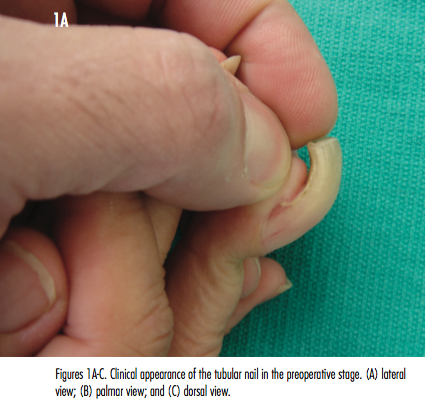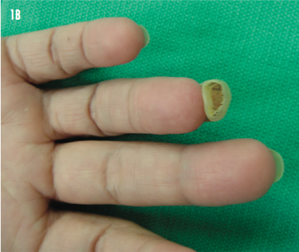
A 56-year-old female presented with a peculiar nail plate shape of the right ring finger that had been present for 15 years, but for the past year had become more bothersome (Figures 1A-C). The patient complained about the mass appearance, noting that its shape and size were making trimming the nail difficult. During this 15-year period, there was a gradual progression of her right ring finger nail plate, from a flat surface to a tubular one. There was no history of trauma or infection to that digit. There was no family history of a similar condition. The rest of her personal and family history was not contributory. On physical examination, the nail plate of the right ring finger had become a raised tube surrounding a nail bed-like tissue approximating 1 cm x 1 cm in size. No other digit was affected. No other skin or mucosal lesions of clinical significance were identified.
What's Your Diagnosis?
To learn the answer, go to page 2
{{pagebreak}}

Diagnosis: Solitary Subungual Neurofibroma of the Right Ring Finger Resulting in a Tubular Nail Plate
Neurofibromas are benign tumors that arise from the peripheral nerves affecting any area of the body. The 3 most common types are classified as cutaneous, spinal, and plexiform. Our patient presented with the cutaneous subtype. We report a case of a solitary subungual neurofibroma occurring in the right ring finger nail bed. To date, there has been only 10 other noted cases, but none forming a tubular nail plate.1
Clinical Presentation
Subungual neurofibromas present as slow growing tumors with onychodystrophy and minimal clinical signs.2,3 However, it has been reported that patients can present with pain in the affected digit. Currently, trends within the literature demonstrate that this disease is not associated with trauma, and also describe a predilection for affecting middle-aged women.4 These latter 2 factors were consistent with our patient’s presentation. Neurofibromas are a rare finding in the digits and when they occur as an isolated lesion with no association with neufibromatosis type 1 (von Recklinghausen disease), the presentation is even more unique. Solitary subungual neurofibromas were first seen and reported by Runne and Orfanoes4 in 1981 and since then only 10  other cases of solitary subungual neurofibromas have been reported.1 The diagnosis of this type of tumor is often not apparent as they are usually indiscrete in size and often lack clinical symptoms. Because of these factors, excision of the tumor is of both diagnostic and therapeutic value.
Histopathology
Histologically these tumors contain a loose array of spindle cells, which have pale cytoplasm and wavy nuclei. In the background is often a myxoid stroma with mast cells seen throughout the neurofibroma. On immunostaining, tumors can be positive for S-100 protein, vimentin, and epithelial membrane antigen.2,4 While the mass is unencapsulated, these subungual tumors are well circumscribed. These features are specific for neurofibromas and help differentiate this tumor from other potential diagnoses. Histology in our patient was consistent with a neurofibroma, with a majority of the parenchymal cells staining positive for the S-100 protein.
Differential Diagnosis
Glomus tumor of the digit has a propensity of being subungual; however, this tumor has hypersensitivity to cold, paroxysmal severe pain, and point tenderness. These are not seen in subungual neurofibromas. Glomus tumors also rarely deform the nail plate. There are also other entities that can present  like subungual neurofibromas. Epidermoid cysts, fibrokeratomas, Koenen tumors, and squamous cell carcinomas are some of the subungual disease processes to be considered in the differential.5
Management
Treatment of choice for patients affected with subungual neurofibromas is always surgical excision. After resection of a subungual neurofibroma, patients can be reassured that reoccurrence has not been reported in literature.5 Most have regained a good cosmetic appearance with the regrowth of the nail plate. Another important aspect in the management of these patients is to determine if there is local skeletal involvement from the tumor. Some cases reported in the literature have shown radiologic evidence of erosion or scalloping of the phalangeal cortex due to the neurofibroma.2
Our Patient
There was no family history of neurofibromatosis type 1 in our patient. On examination, the nail had a tubular shape with a very thin dorsal nail plate and a thick deep nail plate (Figures 1A-C). The undersurface of the nail appeared to be attached to the nail bed, with the nail tissue growing into the hollow tube. The nail near the origin of the growth appeared to be thin to a point where the soft tissue below could be seen. No other signs of neurofibromatosis type 1, such as café-au-lait spots, axillary freckling, or Lisch nodules were noted on examination. An initial x-ray showed no evidence of bony involvement. Elective surgery was then performed. Two axial nail incisions in the nail fold elevating the eponychium allowed exposure of a soft fleshy white dense mass originating from the nail germinal and sterile matrix (Figure 2). This mass was excised flush with the nail bed. Histologic examination confirmed the diagnosis of a solitary subungual neurofibroma. Healing and regrowth of the nail were uneventful on follow-up visits. There was no tumor recurrence at 6 months following surgery.

Conclusion
This case illustrates an unusual tubular nail caused by a solitary subungual neurofibroma. To our knowledge, this peculiar shape of a tubular nail deformity seen in our patient has not been reported in the literature as being associated with a solitary subungual neurofibroma. The fingers and toes have been the most common areas affected in reported cases and middle-aged women seem to be the most vulnerable to the condition.4 These tumors often progress slowly over time and can present with nonspecific symptoms such as pain. The final diagnosis can only be made through excision of the mass followed by histologic examination. Excision is also of therapeutic value with patients having excellent clinical outcomes. To date, there has not been any reported cases of recurrence in the literature.5
Mr Burton is a medical student, MS-4, at the University of Central Florida College of Medicine in Orlando, FL.
Ms Humphries is a medical student, MS-4, at the University of Central Florida College of Medicine in Orlando, FL.
Dr Macksoud is an orthopedic surgeon with Jewett Orthopaedic Clinic in Orlando, FL.
Disclosure: The authors report no relevant financial relationships.
References
1. Huajun J, Wei Q, Ming L, Chongyang F, Weiguo Z, Decheng L. Solitary subungual neurofibroma in the right first finger. Int J Dermatol. 2012;51(3):335-338.
2. Dangoisse C, Andre J, De Dobbeleer G, Van Geertruyden J. Solitary subungual neurofibroma. Br J Dermatol. 2000;143(5):1116-1117.
3. Stolarczuk Dde A, Silva AL, Filgueiras Fda M, Alves Mde F, Silva SC. Solitary subungual neurofibroma: a previously unreported finding in a male patient. An Bras Dermatol. 2011;86(3):569-572.
4. Runne U, Orfanos CE. The human nail: structure, growth and pathological changes. Curr Probl Dermatol. 1981;9:102-149.
5. Roldan-Marin R, Dominguez-Cherit J, Vega-Memije ME, Toussaint-Caire S, Hojyo-Tomoka MT, Dominguez-Soto L. Solitary subungual neurofibroma: an uncommon finding and a review of the literature. J Drugs Dermatol. 2006;5(7):672-674.

A 56-year-old female presented with a peculiar nail plate shape of the right ring finger that had been present for 15 years, but for the past year had become more bothersome (Figures 1A-C). The patient complained about the mass appearance, noting that its shape and size were making trimming the nail difficult. During this 15-year period, there was a gradual progression of her right ring finger nail plate, from a flat surface to a tubular one. There was no history of trauma or infection to that digit. There was no family history of a similar condition. The rest of her personal and family history was not contributory. On physical examination, the nail plate of the right ring finger had become a raised tube surrounding a nail bed-like tissue approximating 1 cm x 1 cm in size. No other digit was affected. No other skin or mucosal lesions of clinical significance were identified.
What's Your Diagnosis?

Diagnosis: Solitary Subungual Neurofibroma of the Right Ring Finger Resulting in a Tubular Nail Plate
Neurofibromas are benign tumors that arise from the peripheral nerves affecting any area of the body. The 3 most common types are classified as cutaneous, spinal, and plexiform. Our patient presented with the cutaneous subtype. We report a case of a solitary subungual neurofibroma occurring in the right ring finger nail bed. To date, there has been only 10 other noted cases, but none forming a tubular nail plate.1
Clinical Presentation
Subungual neurofibromas present as slow growing tumors with onychodystrophy and minimal clinical signs.2,3 However, it has been reported that patients can present with pain in the affected digit. Currently, trends within the literature demonstrate that this disease is not associated with trauma, and also describe a predilection for affecting middle-aged women.4 These latter 2 factors were consistent with our patient’s presentation. Neurofibromas are a rare finding in the digits and when they occur as an isolated lesion with no association with neufibromatosis type 1 (von Recklinghausen disease), the presentation is even more unique. Solitary subungual neurofibromas were first seen and reported by Runne and Orfanoes4 in 1981 and since then only 10  other cases of solitary subungual neurofibromas have been reported.1 The diagnosis of this type of tumor is often not apparent as they are usually indiscrete in size and often lack clinical symptoms. Because of these factors, excision of the tumor is of both diagnostic and therapeutic value.
Histopathology
Histologically these tumors contain a loose array of spindle cells, which have pale cytoplasm and wavy nuclei. In the background is often a myxoid stroma with mast cells seen throughout the neurofibroma. On immunostaining, tumors can be positive for S-100 protein, vimentin, and epithelial membrane antigen.2,4 While the mass is unencapsulated, these subungual tumors are well circumscribed. These features are specific for neurofibromas and help differentiate this tumor from other potential diagnoses. Histology in our patient was consistent with a neurofibroma, with a majority of the parenchymal cells staining positive for the S-100 protein.
Differential Diagnosis
Glomus tumor of the digit has a propensity of being subungual; however, this tumor has hypersensitivity to cold, paroxysmal severe pain, and point tenderness. These are not seen in subungual neurofibromas. Glomus tumors also rarely deform the nail plate. There are also other entities that can present  like subungual neurofibromas. Epidermoid cysts, fibrokeratomas, Koenen tumors, and squamous cell carcinomas are some of the subungual disease processes to be considered in the differential.5
Management
Treatment of choice for patients affected with subungual neurofibromas is always surgical excision. After resection of a subungual neurofibroma, patients can be reassured that reoccurrence has not been reported in literature.5 Most have regained a good cosmetic appearance with the regrowth of the nail plate. Another important aspect in the management of these patients is to determine if there is local skeletal involvement from the tumor. Some cases reported in the literature have shown radiologic evidence of erosion or scalloping of the phalangeal cortex due to the neurofibroma.2
Our Patient
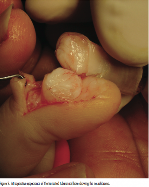
There was no family history of neurofibromatosis type 1 in our patient. On examination, the nail had a tubular shape with a very thin dorsal nail plate and a thick deep nail plate (Figures 1A-C). The undersurface of the nail appeared to be attached to the nail bed, with the nail tissue growing into the hollow tube. The nail near the origin of the growth appeared to be thin to a point where the soft tissue below could be seen. No other signs of neurofibromatosis type 1, such as café-au-lait spots, axillary freckling, or Lisch nodules were noted on examination. An initial x-ray showed no evidence of bony involvement. Elective surgery was then performed. Two axial nail incisions in the nail fold elevating the eponychium allowed exposure of a soft fleshy white dense mass originating from the nail germinal and sterile matrix (Figure 2). This mass was excised flush with the nail bed. Histologic examination confirmed the diagnosis of a solitary subungual neurofibroma. Healing and regrowth of the nail were uneventful on follow-up visits. There was no tumor recurrence at 6 months following surgery.

Conclusion
This case illustrates an unusual tubular nail caused by a solitary subungual neurofibroma. To our knowledge, this peculiar shape of a tubular nail deformity seen in our patient has not been reported in the literature as being associated with a solitary subungual neurofibroma. The fingers and toes have been the most common areas affected in reported cases and middle-aged women seem to be the most vulnerable to the condition.4 These tumors often progress slowly over time and can present with nonspecific symptoms such as pain. The final diagnosis can only be made through excision of the mass followed by histologic examination. Excision is also of therapeutic value with patients having excellent clinical outcomes. To date, there has not been any reported cases of recurrence in the literature.5
Mr Burton is a medical student, MS-4, at the University of Central Florida College of Medicine in Orlando, FL.
Ms Humphries is a medical student, MS-4, at the University of Central Florida College of Medicine in Orlando, FL.
Dr Macksoud is an orthopedic surgeon with Jewett Orthopaedic Clinic in Orlando, FL.
Disclosure: The authors report no relevant financial relationships.
References
1. Huajun J, Wei Q, Ming L, Chongyang F, Weiguo Z, Decheng L. Solitary subungual neurofibroma in the right first finger. Int J Dermatol. 2012;51(3):335-338.
2. Dangoisse C, Andre J, De Dobbeleer G, Van Geertruyden J. Solitary subungual neurofibroma. Br J Dermatol. 2000;143(5):1116-1117.
3. Stolarczuk Dde A, Silva AL, Filgueiras Fda M, Alves Mde F, Silva SC. Solitary subungual neurofibroma: a previously unreported finding in a male patient. An Bras Dermatol. 2011;86(3):569-572.
4. Runne U, Orfanos CE. The human nail: structure, growth and pathological changes. Curr Probl Dermatol. 1981;9:102-149.
5. Roldan-Marin R, Dominguez-Cherit J, Vega-Memije ME, Toussaint-Caire S, Hojyo-Tomoka MT, Dominguez-Soto L. Solitary subungual neurofibroma: an uncommon finding and a review of the literature. J Drugs Dermatol. 2006;5(7):672-674.

A 56-year-old female presented with a peculiar nail plate shape of the right ring finger that had been present for 15 years, but for the past year had become more bothersome (Figures 1A-C). The patient complained about the mass appearance, noting that its shape and size were making trimming the nail difficult. During this 15-year period, there was a gradual progression of her right ring finger nail plate, from a flat surface to a tubular one. There was no history of trauma or infection to that digit. There was no family history of a similar condition. The rest of her personal and family history was not contributory. On physical examination, the nail plate of the right ring finger had become a raised tube surrounding a nail bed-like tissue approximating 1 cm x 1 cm in size. No other digit was affected. No other skin or mucosal lesions of clinical significance were identified.
What's Your Diagnosis?
,

A 56-year-old female presented with a peculiar nail plate shape of the right ring finger that had been present for 15 years, but for the past year had become more bothersome (Figures 1A-C). The patient complained about the mass appearance, noting that its shape and size were making trimming the nail difficult. During this 15-year period, there was a gradual progression of her right ring finger nail plate, from a flat surface to a tubular one. There was no history of trauma or infection to that digit. There was no family history of a similar condition. The rest of her personal and family history was not contributory. On physical examination, the nail plate of the right ring finger had become a raised tube surrounding a nail bed-like tissue approximating 1 cm x 1 cm in size. No other digit was affected. No other skin or mucosal lesions of clinical significance were identified.
What's Your Diagnosis?
To learn the answer, go to page 2
{{pagebreak}}

Diagnosis: Solitary Subungual Neurofibroma of the Right Ring Finger Resulting in a Tubular Nail Plate
Neurofibromas are benign tumors that arise from the peripheral nerves affecting any area of the body. The 3 most common types are classified as cutaneous, spinal, and plexiform. Our patient presented with the cutaneous subtype. We report a case of a solitary subungual neurofibroma occurring in the right ring finger nail bed. To date, there has been only 10 other noted cases, but none forming a tubular nail plate.1
Clinical Presentation
Subungual neurofibromas present as slow growing tumors with onychodystrophy and minimal clinical signs.2,3 However, it has been reported that patients can present with pain in the affected digit. Currently, trends within the literature demonstrate that this disease is not associated with trauma, and also describe a predilection for affecting middle-aged women.4 These latter 2 factors were consistent with our patient’s presentation. Neurofibromas are a rare finding in the digits and when they occur as an isolated lesion with no association with neufibromatosis type 1 (von Recklinghausen disease), the presentation is even more unique. Solitary subungual neurofibromas were first seen and reported by Runne and Orfanoes4 in 1981 and since then only 10  other cases of solitary subungual neurofibromas have been reported.1 The diagnosis of this type of tumor is often not apparent as they are usually indiscrete in size and often lack clinical symptoms. Because of these factors, excision of the tumor is of both diagnostic and therapeutic value.
Histopathology
Histologically these tumors contain a loose array of spindle cells, which have pale cytoplasm and wavy nuclei. In the background is often a myxoid stroma with mast cells seen throughout the neurofibroma. On immunostaining, tumors can be positive for S-100 protein, vimentin, and epithelial membrane antigen.2,4 While the mass is unencapsulated, these subungual tumors are well circumscribed. These features are specific for neurofibromas and help differentiate this tumor from other potential diagnoses. Histology in our patient was consistent with a neurofibroma, with a majority of the parenchymal cells staining positive for the S-100 protein.
Differential Diagnosis
Glomus tumor of the digit has a propensity of being subungual; however, this tumor has hypersensitivity to cold, paroxysmal severe pain, and point tenderness. These are not seen in subungual neurofibromas. Glomus tumors also rarely deform the nail plate. There are also other entities that can present  like subungual neurofibromas. Epidermoid cysts, fibrokeratomas, Koenen tumors, and squamous cell carcinomas are some of the subungual disease processes to be considered in the differential.5
Management
Treatment of choice for patients affected with subungual neurofibromas is always surgical excision. After resection of a subungual neurofibroma, patients can be reassured that reoccurrence has not been reported in literature.5 Most have regained a good cosmetic appearance with the regrowth of the nail plate. Another important aspect in the management of these patients is to determine if there is local skeletal involvement from the tumor. Some cases reported in the literature have shown radiologic evidence of erosion or scalloping of the phalangeal cortex due to the neurofibroma.2
Our Patient
There was no family history of neurofibromatosis type 1 in our patient. On examination, the nail had a tubular shape with a very thin dorsal nail plate and a thick deep nail plate (Figures 1A-C). The undersurface of the nail appeared to be attached to the nail bed, with the nail tissue growing into the hollow tube. The nail near the origin of the growth appeared to be thin to a point where the soft tissue below could be seen. No other signs of neurofibromatosis type 1, such as café-au-lait spots, axillary freckling, or Lisch nodules were noted on examination. An initial x-ray showed no evidence of bony involvement. Elective surgery was then performed. Two axial nail incisions in the nail fold elevating the eponychium allowed exposure of a soft fleshy white dense mass originating from the nail germinal and sterile matrix (Figure 2). This mass was excised flush with the nail bed. Histologic examination confirmed the diagnosis of a solitary subungual neurofibroma. Healing and regrowth of the nail were uneventful on follow-up visits. There was no tumor recurrence at 6 months following surgery.

Conclusion
This case illustrates an unusual tubular nail caused by a solitary subungual neurofibroma. To our knowledge, this peculiar shape of a tubular nail deformity seen in our patient has not been reported in the literature as being associated with a solitary subungual neurofibroma. The fingers and toes have been the most common areas affected in reported cases and middle-aged women seem to be the most vulnerable to the condition.4 These tumors often progress slowly over time and can present with nonspecific symptoms such as pain. The final diagnosis can only be made through excision of the mass followed by histologic examination. Excision is also of therapeutic value with patients having excellent clinical outcomes. To date, there has not been any reported cases of recurrence in the literature.5
Mr Burton is a medical student, MS-4, at the University of Central Florida College of Medicine in Orlando, FL.
Ms Humphries is a medical student, MS-4, at the University of Central Florida College of Medicine in Orlando, FL.
Dr Macksoud is an orthopedic surgeon with Jewett Orthopaedic Clinic in Orlando, FL.
Disclosure: The authors report no relevant financial relationships.
References
1. Huajun J, Wei Q, Ming L, Chongyang F, Weiguo Z, Decheng L. Solitary subungual neurofibroma in the right first finger. Int J Dermatol. 2012;51(3):335-338.
2. Dangoisse C, Andre J, De Dobbeleer G, Van Geertruyden J. Solitary subungual neurofibroma. Br J Dermatol. 2000;143(5):1116-1117.
3. Stolarczuk Dde A, Silva AL, Filgueiras Fda M, Alves Mde F, Silva SC. Solitary subungual neurofibroma: a previously unreported finding in a male patient. An Bras Dermatol. 2011;86(3):569-572.
4. Runne U, Orfanos CE. The human nail: structure, growth and pathological changes. Curr Probl Dermatol. 1981;9:102-149.
5. Roldan-Marin R, Dominguez-Cherit J, Vega-Memije ME, Toussaint-Caire S, Hojyo-Tomoka MT, Dominguez-Soto L. Solitary subungual neurofibroma: an uncommon finding and a review of the literature. J Drugs Dermatol. 2006;5(7):672-674.

A 56-year-old female presented with a peculiar nail plate shape of the right ring finger that had been present for 15 years, but for the past year had become more bothersome (Figures 1A-C). The patient complained about the mass appearance, noting that its shape and size were making trimming the nail difficult. During this 15-year period, there was a gradual progression of her right ring finger nail plate, from a flat surface to a tubular one. There was no history of trauma or infection to that digit. There was no family history of a similar condition. The rest of her personal and family history was not contributory. On physical examination, the nail plate of the right ring finger had become a raised tube surrounding a nail bed-like tissue approximating 1 cm x 1 cm in size. No other digit was affected. No other skin or mucosal lesions of clinical significance were identified.
What's Your Diagnosis?

Diagnosis: Solitary Subungual Neurofibroma of the Right Ring Finger Resulting in a Tubular Nail Plate
Neurofibromas are benign tumors that arise from the peripheral nerves affecting any area of the body. The 3 most common types are classified as cutaneous, spinal, and plexiform. Our patient presented with the cutaneous subtype. We report a case of a solitary subungual neurofibroma occurring in the right ring finger nail bed. To date, there has been only 10 other noted cases, but none forming a tubular nail plate.1
Clinical Presentation
Subungual neurofibromas present as slow growing tumors with onychodystrophy and minimal clinical signs.2,3 However, it has been reported that patients can present with pain in the affected digit. Currently, trends within the literature demonstrate that this disease is not associated with trauma, and also describe a predilection for affecting middle-aged women.4 These latter 2 factors were consistent with our patient’s presentation. Neurofibromas are a rare finding in the digits and when they occur as an isolated lesion with no association with neufibromatosis type 1 (von Recklinghausen disease), the presentation is even more unique. Solitary subungual neurofibromas were first seen and reported by Runne and Orfanoes4 in 1981 and since then only 10  other cases of solitary subungual neurofibromas have been reported.1 The diagnosis of this type of tumor is often not apparent as they are usually indiscrete in size and often lack clinical symptoms. Because of these factors, excision of the tumor is of both diagnostic and therapeutic value.
Histopathology
Histologically these tumors contain a loose array of spindle cells, which have pale cytoplasm and wavy nuclei. In the background is often a myxoid stroma with mast cells seen throughout the neurofibroma. On immunostaining, tumors can be positive for S-100 protein, vimentin, and epithelial membrane antigen.2,4 While the mass is unencapsulated, these subungual tumors are well circumscribed. These features are specific for neurofibromas and help differentiate this tumor from other potential diagnoses. Histology in our patient was consistent with a neurofibroma, with a majority of the parenchymal cells staining positive for the S-100 protein.
Differential Diagnosis
Glomus tumor of the digit has a propensity of being subungual; however, this tumor has hypersensitivity to cold, paroxysmal severe pain, and point tenderness. These are not seen in subungual neurofibromas. Glomus tumors also rarely deform the nail plate. There are also other entities that can present  like subungual neurofibromas. Epidermoid cysts, fibrokeratomas, Koenen tumors, and squamous cell carcinomas are some of the subungual disease processes to be considered in the differential.5
Management
Treatment of choice for patients affected with subungual neurofibromas is always surgical excision. After resection of a subungual neurofibroma, patients can be reassured that reoccurrence has not been reported in literature.5 Most have regained a good cosmetic appearance with the regrowth of the nail plate. Another important aspect in the management of these patients is to determine if there is local skeletal involvement from the tumor. Some cases reported in the literature have shown radiologic evidence of erosion or scalloping of the phalangeal cortex due to the neurofibroma.2
Our Patient
There was no family history of neurofibromatosis type 1 in our patient. On examination, the nail had a tubular shape with a very thin dorsal nail plate and a thick deep nail plate (Figures 1A-C). The undersurface of the nail appeared to be attached to the nail bed, with the nail tissue growing into the hollow tube. The nail near the origin of the growth appeared to be thin to a point where the soft tissue below could be seen. No other signs of neurofibromatosis type 1, such as café-au-lait spots, axillary freckling, or Lisch nodules were noted on examination. An initial x-ray showed no evidence of bony involvement. Elective surgery was then performed. Two axial nail incisions in the nail fold elevating the eponychium allowed exposure of a soft fleshy white dense mass originating from the nail germinal and sterile matrix (Figure 2). This mass was excised flush with the nail bed. Histologic examination confirmed the diagnosis of a solitary subungual neurofibroma. Healing and regrowth of the nail were uneventful on follow-up visits. There was no tumor recurrence at 6 months following surgery.

Conclusion
This case illustrates an unusual tubular nail caused by a solitary subungual neurofibroma. To our knowledge, this peculiar shape of a tubular nail deformity seen in our patient has not been reported in the literature as being associated with a solitary subungual neurofibroma. The fingers and toes have been the most common areas affected in reported cases and middle-aged women seem to be the most vulnerable to the condition.4 These tumors often progress slowly over time and can present with nonspecific symptoms such as pain. The final diagnosis can only be made through excision of the mass followed by histologic examination. Excision is also of therapeutic value with patients having excellent clinical outcomes. To date, there has not been any reported cases of recurrence in the literature.5
Mr Burton is a medical student, MS-4, at the University of Central Florida College of Medicine in Orlando, FL.
Ms Humphries is a medical student, MS-4, at the University of Central Florida College of Medicine in Orlando, FL.
Dr Macksoud is an orthopedic surgeon with Jewett Orthopaedic Clinic in Orlando, FL.
Disclosure: The authors report no relevant financial relationships.
References
1. Huajun J, Wei Q, Ming L, Chongyang F, Weiguo Z, Decheng L. Solitary subungual neurofibroma in the right first finger. Int J Dermatol. 2012;51(3):335-338.
2. Dangoisse C, Andre J, De Dobbeleer G, Van Geertruyden J. Solitary subungual neurofibroma. Br J Dermatol. 2000;143(5):1116-1117.
3. Stolarczuk Dde A, Silva AL, Filgueiras Fda M, Alves Mde F, Silva SC. Solitary subungual neurofibroma: a previously unreported finding in a male patient. An Bras Dermatol. 2011;86(3):569-572.
4. Runne U, Orfanos CE. The human nail: structure, growth and pathological changes. Curr Probl Dermatol. 1981;9:102-149.
5. Roldan-Marin R, Dominguez-Cherit J, Vega-Memije ME, Toussaint-Caire S, Hojyo-Tomoka MT, Dominguez-Soto L. Solitary subungual neurofibroma: an uncommon finding and a review of the literature. J Drugs Dermatol. 2006;5(7):672-674.

Diagnosis: Solitary Subungual Neurofibroma of the Right Ring Finger Resulting in a Tubular Nail Plate
Neurofibromas are benign tumors that arise from the peripheral nerves affecting any area of the body. The 3 most common types are classified as cutaneous, spinal, and plexiform. Our patient presented with the cutaneous subtype. We report a case of a solitary subungual neurofibroma occurring in the right ring finger nail bed. To date, there has been only 10 other noted cases, but none forming a tubular nail plate.1
Clinical Presentation
Subungual neurofibromas present as slow growing tumors with onychodystrophy and minimal clinical signs.2,3 However, it has been reported that patients can present with pain in the affected digit. Currently, trends within the literature demonstrate that this disease is not associated with trauma, and also describe a predilection for affecting middle-aged women.4 These latter 2 factors were consistent with our patient’s presentation. Neurofibromas are a rare finding in the digits and when they occur as an isolated lesion with no association with neufibromatosis type 1 (von Recklinghausen disease), the presentation is even more unique. Solitary subungual neurofibromas were first seen and reported by Runne and Orfanoes4 in 1981 and since then only 10  other cases of solitary subungual neurofibromas have been reported.1 The diagnosis of this type of tumor is often not apparent as they are usually indiscrete in size and often lack clinical symptoms. Because of these factors, excision of the tumor is of both diagnostic and therapeutic value.
Histopathology
Histologically these tumors contain a loose array of spindle cells, which have pale cytoplasm and wavy nuclei. In the background is often a myxoid stroma with mast cells seen throughout the neurofibroma. On immunostaining, tumors can be positive for S-100 protein, vimentin, and epithelial membrane antigen.2,4 While the mass is unencapsulated, these subungual tumors are well circumscribed. These features are specific for neurofibromas and help differentiate this tumor from other potential diagnoses. Histology in our patient was consistent with a neurofibroma, with a majority of the parenchymal cells staining positive for the S-100 protein.
Differential Diagnosis
Glomus tumor of the digit has a propensity of being subungual; however, this tumor has hypersensitivity to cold, paroxysmal severe pain, and point tenderness. These are not seen in subungual neurofibromas. Glomus tumors also rarely deform the nail plate. There are also other entities that can present  like subungual neurofibromas. Epidermoid cysts, fibrokeratomas, Koenen tumors, and squamous cell carcinomas are some of the subungual disease processes to be considered in the differential.5
Management
Treatment of choice for patients affected with subungual neurofibromas is always surgical excision. After resection of a subungual neurofibroma, patients can be reassured that reoccurrence has not been reported in literature.5 Most have regained a good cosmetic appearance with the regrowth of the nail plate. Another important aspect in the management of these patients is to determine if there is local skeletal involvement from the tumor. Some cases reported in the literature have shown radiologic evidence of erosion or scalloping of the phalangeal cortex due to the neurofibroma.2
Our Patient
There was no family history of neurofibromatosis type 1 in our patient. On examination, the nail had a tubular shape with a very thin dorsal nail plate and a thick deep nail plate (Figures 1A-C). The undersurface of the nail appeared to be attached to the nail bed, with the nail tissue growing into the hollow tube. The nail near the origin of the growth appeared to be thin to a point where the soft tissue below could be seen. No other signs of neurofibromatosis type 1, such as café-au-lait spots, axillary freckling, or Lisch nodules were noted on examination. An initial x-ray showed no evidence of bony involvement. Elective surgery was then performed. Two axial nail incisions in the nail fold elevating the eponychium allowed exposure of a soft fleshy white dense mass originating from the nail germinal and sterile matrix (Figure 2). This mass was excised flush with the nail bed. Histologic examination confirmed the diagnosis of a solitary subungual neurofibroma. Healing and regrowth of the nail were uneventful on follow-up visits. There was no tumor recurrence at 6 months following surgery.

Conclusion
This case illustrates an unusual tubular nail caused by a solitary subungual neurofibroma. To our knowledge, this peculiar shape of a tubular nail deformity seen in our patient has not been reported in the literature as being associated with a solitary subungual neurofibroma. The fingers and toes have been the most common areas affected in reported cases and middle-aged women seem to be the most vulnerable to the condition.4 These tumors often progress slowly over time and can present with nonspecific symptoms such as pain. The final diagnosis can only be made through excision of the mass followed by histologic examination. Excision is also of therapeutic value with patients having excellent clinical outcomes. To date, there has not been any reported cases of recurrence in the literature.5
Mr Burton is a medical student, MS-4, at the University of Central Florida College of Medicine in Orlando, FL.
Ms Humphries is a medical student, MS-4, at the University of Central Florida College of Medicine in Orlando, FL.
Dr Macksoud is an orthopedic surgeon with Jewett Orthopaedic Clinic in Orlando, FL.
Disclosure: The authors report no relevant financial relationships.
References
1. Huajun J, Wei Q, Ming L, Chongyang F, Weiguo Z, Decheng L. Solitary subungual neurofibroma in the right first finger. Int J Dermatol. 2012;51(3):335-338.
2. Dangoisse C, Andre J, De Dobbeleer G, Van Geertruyden J. Solitary subungual neurofibroma. Br J Dermatol. 2000;143(5):1116-1117.
3. Stolarczuk Dde A, Silva AL, Filgueiras Fda M, Alves Mde F, Silva SC. Solitary subungual neurofibroma: a previously unreported finding in a male patient. An Bras Dermatol. 2011;86(3):569-572.
4. Runne U, Orfanos CE. The human nail: structure, growth and pathological changes. Curr Probl Dermatol. 1981;9:102-149.
5. Roldan-Marin R, Dominguez-Cherit J, Vega-Memije ME, Toussaint-Caire S, Hojyo-Tomoka MT, Dominguez-Soto L. Solitary subungual neurofibroma: an uncommon finding and a review of the literature. J Drugs Dermatol. 2006;5(7):672-674.


 other cases of solitary subungual neurofibromas have been reported.1 The diagnosis of this type of tumor is often not apparent as they are usually indiscrete in size and often lack clinical symptoms. Because of these factors, excision of the tumor is of both diagnostic and therapeutic value.
  like subungual neurofibromas. Epidermoid cysts, fibrokeratomas, Koenen tumors, and squamous cell carcinomas are some of the subungual disease processes to be considered in the differential.5

 other cases of solitary subungual neurofibromas have been reported.1 The diagnosis of this type of tumor is often not apparent as they are usually indiscrete in size and often lack clinical symptoms. Because of these factors, excision of the tumor is of both diagnostic and therapeutic value.
  like subungual neurofibromas. Epidermoid cysts, fibrokeratomas, Koenen tumors, and squamous cell carcinomas are some of the subungual disease processes to be considered in the differential.5


