Study: PRP As Effective As Steroids For Plantar Fasciitis
 Platelet-rich plasma (PRP) can be an effective treatment for plantar fasciitis in patients who have failed conservative therapy, according to a new study.
The randomized study published recently online by the Journal of the American Podiatric Medical Association focused on 14 patients with plantar fasciitis, who were treated with either intralesional steroids or PRP. Platelet-rich plasma treatment consisted of 3 mL of plasma activated with 0.45 mL of 10% calcium gluconate while steroid treatment consisted of 8 mg of dexamethasone plus 2 mL of lidocaine. Researchers noted that both groups experienced improvements in the Visual Analogue Scale score but differences between groups were not statistically significant. The study authors conclude that PRP is equally effective as steroids for patients with plantar fasciitis but they cite cost and preparation time as disadvantages for PRP.
Lawrence Oloff, DPM, FACFAS, cites “mediocre” results with PRP for plantar fasciitis, noting that he has had better success using the modality for tendon disease. He chooses PRP for heel pain if a patient has failed traditional treatment protocols such as physical therapy, non-steroidal anti-inflammatory drugs, orthotics, night splints or steroid injections. One may consider PRP if the patient is not quite ready to proceed to surgery and is looking for another option, according to Dr. Oloff, who is in private practice at Sports Orthopedic and Rehabilitation Medicine Associates in Redwood City, Calif.
Patrick DeHeer, DPM, FACFAS, has not used PRP for plantar fasciitis as he has found the the literature to be inconclusive on its efficacy. As he notes, most patients with plantar fasciitis respond well to standard protocols including inflammation reduction, biomechanical control and stretching of the gastrocsoleus complex/plantar fascia. For that small group of patients who do not respond, Dr. DeHeer opts for gastrocnemius recessions.
“Platelet-rich plasma is more expensive, time consuming and is not any more effective than steroid injections,” says Dr. DeHeer, who is in private practice with various offices in Indianapolis and is the founder of Step by Step Haiti. “Therefore, I have not discovered adequate evidence-based medicine to support including it in my practice.”
Dr. Oloff cites advantages of PRP including that it does not cause any known ill effects on tissue whereas steroids can degrade tissue over time. However, he notes the advantage to steroids is that they have a longer track record in terms of application for plantar fasciitis.
Editor’s note: For further reading, see “Current Insights On PRP And Achilles Tendinopathy” on page 60 of this month’s issue, “Point-Counterpoint: Is PRP Beneficial For Chronic Plantar Fasciitis?” in the June 2013 issue or “Platelet-Rich Plasma: Can It Have An Impact For Plantar Fasciitis?” in the November 2012 issue of Podiatry Today.
Should Surgeons Use Unmodified K-Wires?
By Brian McCurdy, Managing Editor
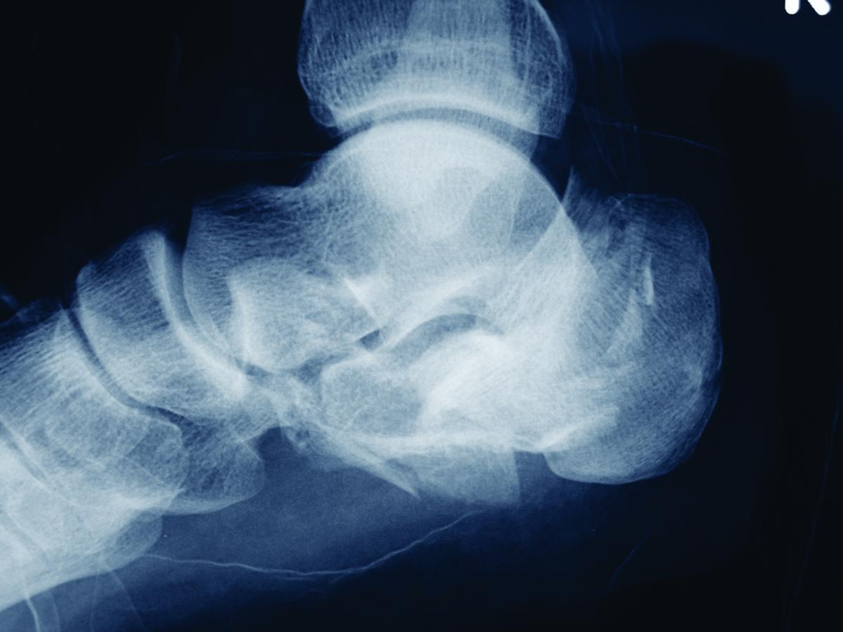
 A recent study in the Journal of Foot and Ankle Surgery discourages the use of unmodified Kirschner wires (K-wires) for open reduction and internal fixation (ORIF) of calcaneal fractures, pointing out an incidence of dislocation and migration with the fixation.
Researchers analyzed data from a total of 279 patients who received surgery for displaced intra-articular calcaneal fractures over a 15-year period. The study identified 69 K-wires that surgeons used in 49 patients, finding that a total of 25 K-wires had been lost (buried), 38 had been bent, and six were unmodified straight K-wires. Overall, authors say secondary dislocation occurred with four out of 69 cases in which surgeons used K-wires and K-wire migration occurred in 5.8 percent of the cases. In contrast, the study notes none of the bent K-wires migrated or led to secondary dislocation in the present study.
William Fishco, DPM, FACFAS, cites several pros to using K-wires for calcaneal fractures. He says they are simple to use, less time-consuming, necessitate less dissection and can facilitate a minimally invasive technique for less complicated fractures. On the downside, he says K-wires are less stable, offer no compression and are unable to pull bone together for sustentaculum fractures.
In his experience with calcaneal fractures, complications of K-wires include migration and breakage as well as lack of anatomic restoration of the posterior facet, according to Dr. Fishco, a faculty member of the Podiatry Institute, who is in private practice in Phoenix. He notes other alternatives to K-wires include mini external fixators for a minimally invasive technique.
“I am personally a strong advocate of AO fixation with screws and a plate for calcaneal fractures,” says Dr. Fishco.
Study Examines Simulated Weightbearing Lateral Imaging For Lapidus Midfoot Fusions
By Brian McCurdy, Managing Editor
Acknowledging skepticism about established methods to determine the sagittal plane relationship of the first ray to the second ray in Lapidus midfoot fusion, a recent study in the Journal of Foot and Ankle Surgery focuses on intraoperative simulated weightbearing lateral foot imaging studies as an effective alternative.
The study authors used a consistent simulated weightbearing technique for 50 consecutive patients who had a Lapidus midfoot fusion in order to achieve parallel sagittal plane alignment of the first and second metatarsals with no divergence. Forty-seven patients had no divergence and three had divergence with mild first ray elevatus. The researchers noted a direct correlation between the intraoperative simulated and postoperative full weightbearing images for all 50 patients. The study authors concluded that findings from the intraoperative imaging technique are a reliable predictor of first ray sagittal plane alignment in Lapidus midfoot fusion.
One of the goals of the study was determining if the intraoperative findings match what is visible postoperatively, notes lead study author Troy Boffeli, DPM, FACFAS. He says this technique also allows consistent imaging, which he feels “can only lead to better results.”
Surgeons commonly use a flat object like a lid from a screw set to evaluate intraoperative foot alignment, according to Dr. Boffeli, the Director of the Foot and Ankle Surgical Residency Program at Regions Hospital/HealthPartners Institute for Education and Research in St. Paul, MN. Although X-rays are the most accurate way to assess first ray position in comparison to the second ray, he notes X-rays are only useful if the view is from the correct lateral position. Dr. Boffeli says the common technique of loading the foot by using an Army–Navy retractor or the wide end of a mallet to load the forefoot is effective, but does not put all of the metatarsal heads on the same plane.
Dr. Boffeli uses the intraoperative simulated weightbearing technique for almost all reconstructive foot and ankle procedures in which it is important on lateral imaging to assess medial column and rearfoot alignment. He commonly obtains the initial simulated weightbearing lateral foot image after placing temporary fixation and obtains comparative imaging after permanent fixation. He finds the simulated weightbearing technique to be useful for dorsiflexory wedge osteotomies, Cotton osteotomies, Evans osteotomies, ankle arthrodesis, subtalar arthrodesis, naviculocuneiform arthrodesis and talonavicular arthrodesis.
“Foot and ankle surgeons are very astute at examining lateral radiographs regarding first ray alignment, Meary’s angle, talocalcaneal alignment, etc.,” says Dr. Boffeli. “We rely on accurate lateral imaging to assess these parameters. Therefore, proper lateral imaging in the operating room makes sense.”


