ADVERTISEMENT
Study Compares Proximal Chevron And Double Metatarsal Osteotomies For Hallux Valgus
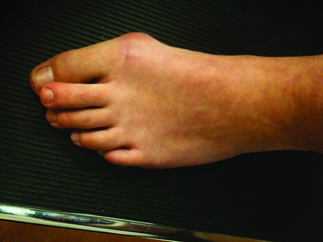
 A recent study found no differences in the clinical and radiographic results following proximal chevron osteotomies and double metatarsal osteotomies in patients with hallux valgus with an increased distal metatarsal articular angle.
The study, published in the Journal of Foot and Ankle Surgery, focused on a proximal chevron osteotomy with Akin osteotomy and lateral soft tissue release in 46 feet, and a double metatarsal osteotomy and Akin osteotomy without lateral soft tissue release in 23 feet. The authors noted the immediate postoperative hallux valgus angle and sesamoid position were significantly larger in the double metatarsal osteotomy group. This group also had a smaller postoperative distal metatarsal articular angle. In addition, the mean shortening of the first metatarsal after surgery was significantly larger in the double metatarsal osteotomy group than in the proximal chevron osteotomy group.
Lawrence Fallat, DPM, FACFAS, utilizes several procedures to reduce a high distal metatarsal articular angle (greater than 9 or 10 degrees). He will perform a distal transverse osteotomy in the metatarsal head with the base of the wedge facing medially. Dr. Fallat says the procedure is technically easy to perform and by increasing the size of the medial wedge, it is easy to accurately correct the distal metatarsal articular angle. Previously, he would make the osteotomy just proximal to the articular surface of the first metatarsal but found fixation challenging. Now Dr. Fallat will make the osteotomy more proximal but still in the metatarsal head, leaving the lateral cortex intact and using a staple or a K-wire for stability.
For patients with hallux valgus and an increased distal metatarsal articular angle, Danielle Butto, DPM, prefers to use the Lapidus procedure with triplane correction. She has found that reduction of the frontal plane, correction of the intermetatarsal angle in the transverse plane and realignment of the first metatarsal in the sagittal plane adequately addresses the deformity present at the first metatarsophalangeal joint (MPJ). She does not perform a lateral release or a medial eminence resection.
Based on the study results, Dr. Butto does not see an advantage to performing a double osteotomy. Disadvantages of the double osteotomy include two osteotomy sites that need to heal, avascular necrosis, first metatarsal shortening and the creating of two new center of rotation of angulation (CORA) points, notes Dr. Butto, an Attending Physician and the Director of Podiatric Residency Research at St. Francis Hospital and Medical Center in Hartford, Conn.
Dr. Fallat also uses a distal chevron osteotomy with a long dorsal arm. As he notes, one can rotate the head of the metatarsal medially and perform bicortical fixation through the long dorsal arm using one or two screws. This technique can minimize shortening of the metatarsal, according to Dr. Fallat, the Program Director of the foot and ankle surgical residency at Beaumont Hospital–Wayne in Wayne, Mich.
Dr. Butto cites several advantages of the proximal chevron osteotomy in comparison to the double metatarsal osteotomy: theoretically less shortening, only one osteotomy site to heal and correction of the deformity that is more proximal and closer to the CORA, which is present at the first tarsometatarsal joint.
As Dr. Butto notes, disadvantages include the need to perform the osteotomy in close proximity to the communicating artery between the dorsal and plantar first metatarsal vasculature. Dr. Butto says this can lead to disruption of the blood supply as the osteotomy creates a new CORA.
A Closer Look At A New ACFAS Consensus Statement On Infracalcaneal Heel Pain
By Brian McCurdy, Managing Editor
The American College of Foot and Ankle Surgeons has released a new consensus statement that examines the diagnosis of infracalcaneal heel pain and numerous treatments.
Among the consensus panel’s findings, in press in the Journal of Foot and Ankle Surgery, are that plantar fasciitis diagnosis usually depends on history and physical exams alone, radiographs are not routinely necessary to diagnose plantar fasciitis, and advanced imaging techniques such as magnetic resonance imaging (MRI) and ultrasound are not necessary for diagnosis.
Nicholas Romansky, DPM, FACFAS, emphasizes the importance of a thorough diagnosis of heel pain, including examining patients with the hands and doing a baseline X-ray. Although he does not think advanced imaging is necessary, if the heel pain does not improve, MRI may be useful, according to Dr. Romansky, who is in private practice in Media and Phoenixville, Pa.
In regard to treatment, the panel found that stretching, corticosteroid injections and extracorporeal shockwave therapy (ESWT) are safe and effective for plantar fasciitis. For chronic, refractory plantar fasciitis, the panel found safe and effective treatments to include plantar fasciotomy and gastrocnemius release. In contrast, the panel found the evidence on several plantar fasciitis treatments to be uncertain. These treatments included non-steroidal anti-inflammatory drugs (NSAIDs), platelet-rich plasma, amniotic tissue, botulinum toxin, needling, prolotherapy, cryosurgery and bipolar radiofrequency ablation.
As for treatment of heel pain, Dr. Romansky says physicians should teach patients to stretch correctly as he has had patients present who stretched the wrong way. Dr. Romansky notes cortisone injections work well if the heel pain is truly plantar fasciitis. He has found that ESWT works as an adjunct to other treatments.
Dr. Romansky also stresses educating patients about heel pain, saying many present after visiting “Dr. Google and Dr. Amazon.” He also advocates that sedentary patients get up every 20 minutes and walk 20 yards to help prevent heel pain.
Are X-Rays Or MRIs Better Diagnostic Indicators For Osteomyelitis?
By Brian McCurdy, Managing Editor
 Radiographs are superior to magnetic resonance imaging (MRI) in diagnosing osteomyelitis, according to a recent study in Foot and Ankle Specialist.
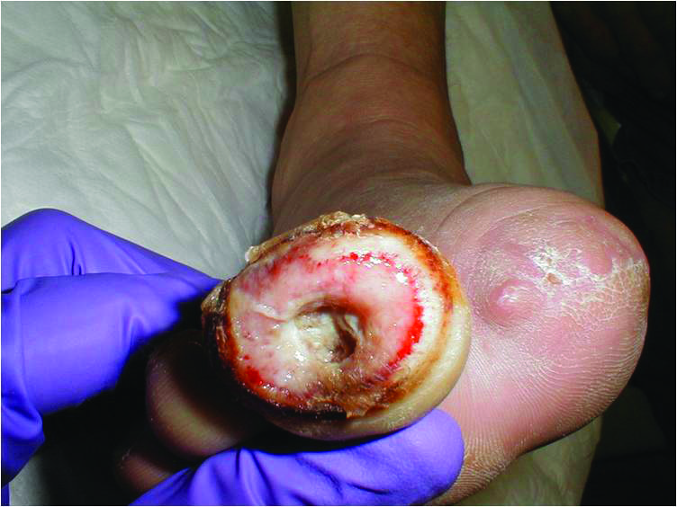
The study focused on 107 patients who had first-time partial foot amputations with confirmed osteomyelitis via histopathological specimens. The authors concluded that plain radiographs were significantly more powerful than MRI in predicting and diagnosing diabetic foot osteomyelitis. The study also confirmed higher erythrocyte sedimentation rate (ESR) values were more likely to reveal a positive diagnosis of osteomyelitis. Researchers noted that intraoperative bone cultures that positively identified bacterial formation did not always indicate true osteomyelitis on histopathological examination.
For Michael Theodoulou, DPM, bone biopsy with culture remains the gold standard for diagnosing osteomyelitis. He adds that multiple site biopsy is frequently recommended so as not to potentially miss infection.
As for imaging techniques, Dr. Theodoulou notes MRI is “the most sensitive imaging we currently have available in the diagnosis of osteomyelitis.” However, he cites a risk of a false positive on MRI, noting other disease states such as neurogenic osteodystrophy or Charcot may present like osteomyelitis on MRI.
Dr. Theodoulou notes plain film X-rays can provide a gross sense of osseous destructive process associated with infection. Although physicians most often use X-rays as a screening tool for osteomyelitis, he will rely on X-rays alone if there is open wound with a “positive probe to bone test” when dealing with phalanges of toes. In the presence of this clinical finding with cardinal signs of infection including redness, swelling, drainage, malodor and/or pain, bone destructive changes as visible on plain film X-rays are highly suggestive of contiguous osteomyelitis, according to Dr. Theodoulou, an Instructor of Surgery at Harvard Medical School.
“In these cases, I cannot justify the cost of a MRI study, which is another disadvantage of MRI,” notes Dr. Theodoulou.
In general, Dr. Theodoulou initially evaluates all suspected cases of osteomyelitis with plain film X-rays and laboratory studies. If his suspicion of osteomyelitis remains high, he then frequently orders MRI to evaluate the extent of the disease process. Ultimately, he will proceed with definitive bone biopsy and culture or a curative-type procedure through debridement of infected bone or limited amputations, and any associated medical management with culture-driven antibiotics.
“Osteomyelitis can be a challenging diagnosis but there are clinical findings, laboratory and radiologic tests that can help provide a more accurate diagnosis,” says Dr. Theodoulou.


