ADVERTISEMENT
Preventing Complications Of Plantar Plate Repair
Sharing insights from their experience as well as the emerging literature on plantar plate surgery, these authors examine current controversies over the alleged association between second metatarsal length and plantar plate pathology, review the benefits of the dorsal approach and offer step-by-step pearls to help thwart potential complications.
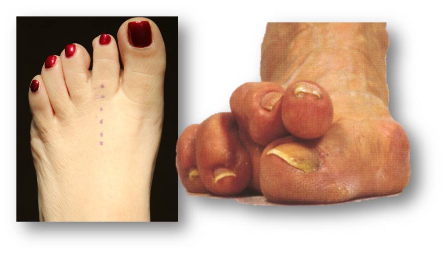
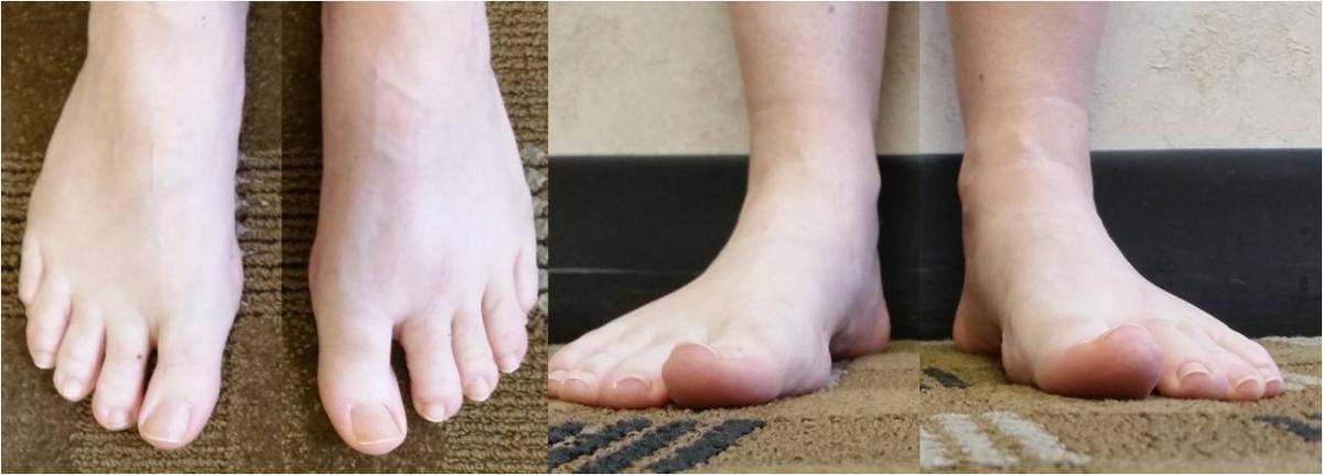
Plantar plate pathology continues to be a controversy in forefoot surgery. Although many surgeons agree that this structure can cause problems and pain, this is not a universally accepted truth. Additionally, for surgeons who do repair this structure, the technique and instrumentation they utilize may be different and outcomes may vary from surgeon to surgeon.
 In order to best address complications related to plantar plate repair, it is important to review what makes this procedure successful.
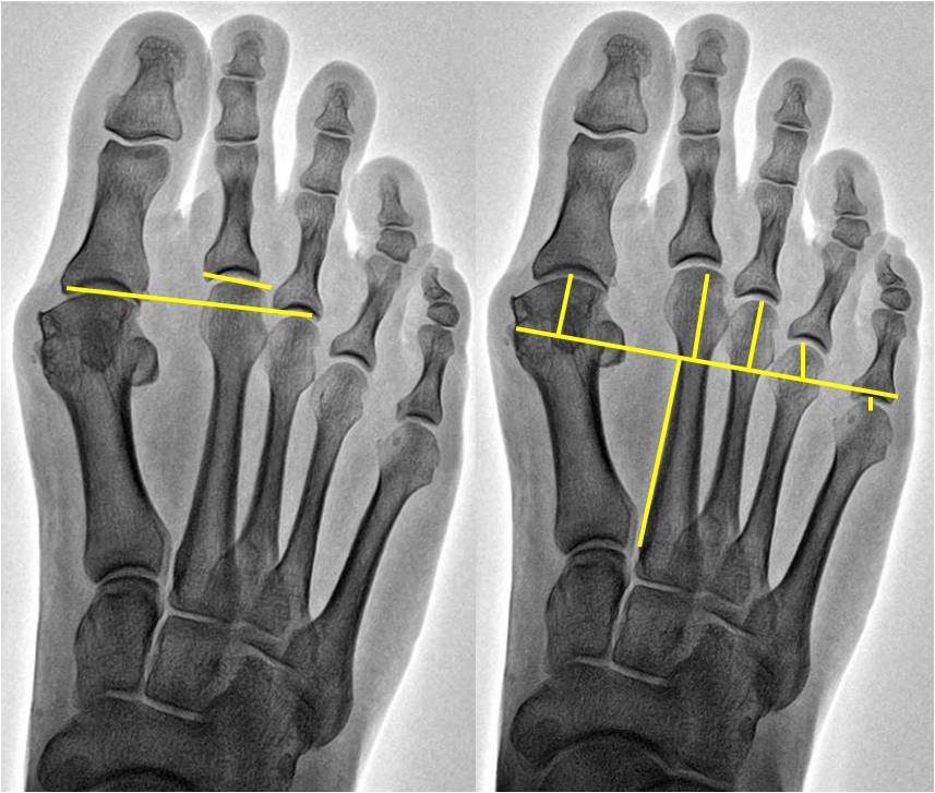
While the metatarsal parabola is an important part of plantar plate repair, there is not a strong understanding of what constitutes an “optimal” parabola. Sansone described the metatarsal parabola as a source of forefoot imbalance and possible source of pain.1 Over time, many authors have tried to describe the “harmonious” parabola or the “correct parabola.”2-4 This has resulted in multiple ways of measuring the metatarsal parabola and more recently, authors have questioned the reliability of measurement of these structures.5,6
One of the current controversies in plantar plate repair—and a point that may be related to the overall success of this procedure—is performing a Weil osteotomy at the same time as the plantar plate repair. In the initial description of a dorsal approach plantar plate repair, Weil and colleagues utilize a single dorsal incision and a Weil metatarsal osteotomy to access the plantar plate.7 After the repair, surgeons pulled the capital fragment of the osteotomy out to the corrected length. This generally shortened the second metatarsal 2 to 3 mm, which corrected the elongated second metatarsal that was so often associated with plantar plate pathology.8
This has led to debate in the forefoot surgery community as many surgeons do not feel one needs to correct the length of the second metatarsal. The thought process is that this is unnecessary as a plantar plate pathology is “a soft tissue problem.” When examining other “soft tissue problems” in the foot and ankle, however, surgeons frequently use osseous correction in conjunction with soft tissue repair to correct the underlying deformity and protect the soft tissue repair. This is the case for a posterior tibial tendon tear that occurs in conjunction with an adult-acquired flatfoot deformity as well as a lateral ankle instability or peroneal tendon pathology that occurs with a cavus foot deformity.9-11
Why is the forefoot different? Frankly, it is not.
A Closer Look At Conflicts In The Literature Over Metatarsal Length
There have been several authors who have argued that second metatarsal length has no relationship to plantar plate pathology.5,9,12,13 Specifically, Kaipel and colleagues found no correlation between the relative length of the first and third metatarsals and maximal peak pressure or maximal force under the corresponding metatarsal heads.9 They noted that relative metatarsal length has no influence on the parameters of plantar loading.
In a study looking at plantar pressure in 12 cadavers after Weil or chevron osteotomies of the second metatarsal, Snyder and colleagues found no significant effect on load in the forefoot after a Weil osteotomy.13 While the authors noted a significant load increase in the first metatarsal region and significant decreases in load under the operated metatarsal heads after the chevron osteotomy, they asserted that the load and pressure changes “occurred independently.” They also maintained that there were no significant changes in the non-operated metatarsal regions.
There have been authors who have argued that the length of the second metatarsal is related to plantar plate pathology.14-16 For example, Bhutta and coworkers found a positive correlation between the length of the second metatarsal and second MPJ instability in 81 patients.14 In 106 feet in 88 patients, Klein and colleagues compared the metatarsal parabola of the foot with symptomatic plantar plate pathology to that of the asymptomatic foot, finding the symptomatic foot had a significantly increased second metatarsal protrusion distance.15
Waverly and coworkers addressed this topic in a study presented at the American College of Foot and Ankle Surgeons (ACFAS) Annual Scientific Conference this past year.17 One hundred total feet were included in this study. In order to be included, patients had to have had surgery on the second metatarsophalangeal joint (MPJ) on one foot by a surgeon from our practice and had no surgery on the contralateral foot. Patients had pressure analysis on both feet and bilateral weightbearing radiographs. The radiographic analysis focused on evaluation of the metatarsal parabola by four different methods. Researchers then correlated this to the pressure analysis data and performed multiple linear regression analyses.
There was a significant relationship between the first and second metatarsals.17 Increased distance between the first and second metatarsal heads was associated with an increase in the ratio of peak pressure between the first and second metatarsal heads. This relationship was statistically significant and maintained in a multiple linear regression analysis.
The absolute length of the second metatarsal and the length of the second metatarsal relative to the third metatarsal were both associated with increases in peak pressure under the second metatarsal head.17 Neither relationship was maintained when the linear regression models controlled for the weight of the patient.
Finally, the length of the second metatarsal had a positive association with the load accepted during ambulation.17 This finding would suggest that as the length of the second metatarsal increases, so does the applied load during gait. Extrapolating this data then would suggest that increasing the load on the second MPJ over time may lead to a repetitive pressure loading phenomenon, which could lead to attenuation of the plantar plate and subsequent tearing of this structure.
Fleischer and colleagues published a second study in 2017 that was a case control-matched trial.7 There were 300 total patients and two controls for each case. The authors defined a case as involving a patient who had a non-acute, isolated second MPJ pain with intraoperative confirmation of a plantar plate injury. Controls were patients who had pain outside the forefoot but were matched in age, gender and year of presentation. The authors reviewed medical records for demographic information, body mass index and smoking status. Researchers reviewed radiographs for forefoot angles and metatarsal parabola measurements.
 The study authors found that the only variable that had an association with plantar plate pathology on the univariate and multivariate analysis was a metatarsal protrusion distance of greater than 4 mm.7 Patients with a second metatarsal length greater than 4 mm had a nearly 2.5 times greater chance of plantar plate injuries. Furthermore, an elongated second metatarsal exhibited a positive dose–response relationship. Patients with increasingly longer second metatarsals were more likely to present with plantar plate injuries.
Although both studies have many good findings, they were both single-site, retrospective studies and the study patients were primarily female.7,17 That being said, however, there is clearly good evidence now suggesting that an abnormal metatarsal parabola is a risk factor for plantar plate injury.
How The Dorsal Approach Enables Easier Intraoperative Identification Of Pathology
The dorsal approach to plantar plate repair allows access to all of the joint structures. A surgeon can visualize the plantar plate, the collateral and accessory ligaments, the metatarsal head, the base of the proximal phalanx and the flexor tendons. If a neuroma is present, that will be easily visible as well. One can address all the pathology from this perspective. If the collateral ligaments are torn, one can repair them at the same time as the plantar plate. If there is an osteochondral lesion or other osseous/cartilaginous component to the problem, surgeons can correct this as well.
When dissecting this region, there are a few things to keep in mind. First, be mindful of the collateral ligament structures. Dissecting the collateral ligaments from the base of the proximal phalanx protects the arterial blood supply to the metatarsal head. The small blood vessels that feed the metatarsal head are not easily visible in vivo but do course near the collateral attachment to the metatarsal head.
Dissection of the plantar plate from the base of the proximal phalanx should occur in a single plane to create a thick flap of tissue. In doing so, the scalpel will come very close to the flexor tendons. The flexor tendons need to remain intact and the plantar plate needs to be in one piece. This will allow a thicker flap of tissue to repair. The location of the plantar plate and flexor tendons can vary, particularly if a collateral ligament is torn or if the toe has changed position quite a bit. In some cases, initial inspection may yield concern about the apparent lack of tissue to repair. Many times, we have found the plantar plate has migrated either medially or laterally on the metatarsal head. There have been a few cases in which the flexor tendons were dislocated and located in the first interspace. These repairs are significantly harder and generally yield less optimal results.
One of the most common complications of this procedure is postoperative arthrofibrosis. This is defined as lack of motion at the joint that is secondary to fibrosis of the joint. This occurrence of this phenomenon is not well understood. Plausible explanations include lack of adherence to the postoperative bracing protocol; physical therapists who are not familiar with the procedure and who are unsure as to how to facilitate the post-op rehab; the biopsychosocial makeup of the patient; the patient’s fear of moving the toe postoperatively; and/or the genetic makeup of the patient. Current investigations are ongoing to understand this complication better.
 Does The Dorsal Approach For Plantar Plate Repair Restore Foot Function And Decrease Pain?
Yes. It does. Two recent studies, one of which is in press at this time, shed some light on this issue.
In the first study, which was presented at the aforementioned ACFAS Annual Scientific Conference, researchers identified 53 patients who had an isolated second metatarsophalangeal joint plantar plate repair by one of two surgeons who utilized the same technique and postoperative protocol.18 The outcomes measures were the Visual Analog Scale (VAS) for pain and the Foot and Ankle Outcome Score. The research team identified that the VAS rating for pain decreased from an average of 6.5 preoperatively to an average of 1.5 postoperatively. The Foot and Ankle Outcome Score significantly improved on four of the five subscales with the fifth subscale trending toward but not obtaining statistical significance. On the physical exam, the patients had alleviation of pain and restoration of stability to the second MPJ.
Flint and colleagues recently published a dorsal approach outcomes study.8 This is a prospective case series of 138 plantar plate repairs in 97 patients. Eighty percent of patients had good to excellent results. The VAS rating for pain decreased from 5.4 to 1.4. The American Orthopaedic Foot and Ankle Society (AOFAS) mean score increased from 49 to 81.
In Conclusion
There are a few key points that one should take away from this discussion:
• be sure to be clear on your technique;
• identify and then correct all noted intraoperative pathology; and
• recognize the importance of the metatarsal parabola.
In summary, the dorsal approach to plantar plate repair is a procedure that can correct pathology of the lesser MPJ while decreasing pain and restoring function to the area.
Dr. Klein is the Associate Director of Research at the Weil Foot, Ankle and Orthopedic Institute. She is a Fellow of the American College of Foot and Ankle Surgeons and a Diplomate of the American Board of Podiatric Medicine.
Dr. Weil is the President of the Weil Foot and Ankle Institute. He is a Fellow of the American College of Foot and Ankle Surgeons.
References
- Sansone S. Forefoot imbalance due to alteration of metatarsus parabola. J Natl Assoc Chirop. 1957; 47(5):219-27.
- Chauhan D, Bhutta M, Barrie J. Does it matter how we measure metatarsal length? Foot Ankle Surg. 2011; 17(3):124-7.
- Chilvers M, Manoli A. The subtle cavus foot and association with ankle instability and lateral foot overload. Foot Ankle Clin. 2008; 13(2):315-24.
- Deben SE, Pomeroy GC. Subtle cavus foot: diagnosis and management. J Am Acad Orthop Surg. 2014; 22(8):512-20.
- Devos Bevernage B, Leemrijse T. Predictive value of radiographic measurements compared to clinical examination. Foot Ankle Int. 2008; 29(2):142-9.
- Domínguez-Maldonado G, Munuera-Martinez P, Castillo-López J, et al. Normal values of metatarsal parabola arch in male and female feet. ScientificWorldJournal. 2014; 6; 2014:505736.
- Fleischer A, Klein E, Ahmad M, et al. Association of abnormal metatarsal parabola with second metatarsophalangeal joint plantar plate pathology. Foot Ankle Int. 2017; 38(3):289-297.
- Flint W, Macias D, Jastifer J, Doty J, Hirose C, Coughlin M. Plantar plate repair for lesser metatarsophalangeal joint instability. Foot Ankle Int. 2017; 38(3):234-242.
- Kaipel M, Krapf D, Wyss C. Metatarsal length does not correlate with maximal peak pressure and maximal force. Clin Orthop Relat Res. 2011; 469(4):1161-6.
- Kaz A, Coughlin M. Crossover second toe: demographics, etiology, and radiographic assessment. Foot Ankle Int. 2007; 28(12):1223-37.
- Khalafi A, Landsman A, Lautenschlager E, Kelikian A. Plantar forefoot pressure changes after second metatarsal neck osteotomy. Foot Ankle Int. 2005; 26(7):550-5.
- Melamed E, Schon L, Myerson M, Parks M. Two modifications of the Weil osteotomy: analysis on sawbone models. Foot Ankle Int. 2002; 23(5):400-5.
- Snyder J, Owen J, Wayne J, Adelaar R. Plantar pressure and load in cadaver feet after a Weil or chevron osteotomy. Foot Ankle Int. 2005; 26(2):158-65.
- Bhutta M, Chauhan D, Zubairy A, Barrie J. Second metatarsophalangeal joint instability and second metatarsal length association depends on the method of measurement. Foot Ankle Int. 2010; 31(6):486-91.
- Klein E, Weil L, Weil L, Knight J. The underlying osseous deformity in plantar plate tears: a radiographic analysis. Foot Ankle Spec. 2013; 6(2):108-18.
- Pinney S, Lin S. Current concept review: acquired adult flatfoot deformity. Foot Ankle Int. 2006; 27(1):66-75.
- Waverly B, Haller S, Weil L, et al. Increased 2nd metatsrsal length is associated with increased peak pressures beneath the 2nd metatarsal head on plantar pressure analysis. Foot Ankle Int. In press.
- Klein E, Weil L, Weil L, Fleischer A. Intermediate term outcomes of the dorsal approach in plantar plate repair. Foot Ankle Spec. In press.
- Weil L, Sung W, Weil L, Malinoski K. Anatomic plantar plate repair using the Weil metatarsal osteotomy approach. Foot Ankle Spec. 2011; 4(3):145-50.
- Maestro M, Besse J, Ragusa M, Berthonnaud E. Forefoot morphotype study and planning method for forefoot osteotomy. Foot Ankle Clin. 2003; 8(4):695-710.


