Is Microfracture Obsolete For Osteochondral Lesions Of The Talus?
 An osteochondral lesion of the talus is often a difficult problem to treat. There is limited understanding of cartilage damage and its repair. Furthermore, it is hard to figure out why some osteochondral lesions, small or large, are painful and some are not painful. Finally, the treatment of such lesions is comprehensive but not standardized. Over time, treatment options have improved and led to better outcomes, but there is no clear and concise understanding of osteochondral lesion treatment, namely what surgeons do and why.
Accordingly, I would like to discuss some of the latest thoughts and ideas on osteochondral lesion treatment, and what I have made my standards of treatment for different lesions.
To understand the treatment options for osteochodral lesions of the talus and the logic behind treatments, it is most important to understand the mechanics of the injury and how different types of lesions can require different treatments. We well understand that the main cause of an osteochondral lesion of the talus is an injury to the cartilage and possibly to the bone. This is mainly either due to a forced stress on the talus or a rotational injury such as a severe ankle sprain. The injury forces the talus into a non-anatomical position, resulting in a shear or crush force that damages the cartilage and bone, causing the underlying injury.
When implementing a treatment regimen, the main points that are necessary to consider in grading and treating a lesion are the size, the depth and the level of underlying bone damage. The logic behind considering the size, depth and underlying bone damage is simple. The larger the lesion, the more aggressive you need to be with your cartilage repair treatments. The deeper the lesion, the more likely you will need to augment the cartilage with bone graft or fill a lesion under the cartilage with some form of a bone substitute.
Finally, if there is significant bone marrow edema, one should consider the subchondral bone injury at the time of cartilage and bone repair. In over 20 years of treating over 500 cartilage lesions, I have never reattached a loose piece of cartilage with screws or pins.
Diagnostic Insights For Osteochondral Lesions
At our institute, we concentrate mainly on the size of lesion as the primary issue. The bigger the size, the more aggressive we are with treatment. We secondly deal with the depth of the lesion. Do we need to graft the lesion? Finally, we deal with the subchondral bone and edema. That’s it. It is that simple.
However, these patients may need subtle adjustments to their treatment. For example, what if you have a small chondral lesion but a large subchondral lesion? What if you have a cartilage damage and the underlying cystic lesion is very large at about 2 cm? These subtle issues require some treatment adjustment but we still go back to considering the size, depth and subchondral bone quality after all is said and done.
Workup of these lesions starts with a history and radiographic examination. Often, the patient has had an injury to the ankle and has had a period of several months to years of conservative care with a chronic ache in the ankle. The patient may report associated periods of locking or catching of the ankle joint. There may also be pain with range of motion although this is rare. Patients will often report a deep ankle pain.
 Radiographs are of very minimal use for osteochondral lesions but can sometimes show a cystic lesion or a flecked piece of cartilage. Our standard workup involves a magnetic resonance image (MRI) for an initial diagnosis after taking radiographs. We prefer MRI over a computed tomography (CT) scan as MRI allows workup of the ligaments, tendons, cartilage, bone and subchondral bone. An MRI is a near complete examination and provides a great deal of necessary information. We concentrate on the size of the lesion, the depth of the lesion and the quality and edema in the subchondral bone. If there is a fairly small lesion with minimal bone involvement and minimal subchondral edema, we know all we need to know.
However, what often goes overlooked with larger lesions is the fact that some of the edema may be causing the lesion to appear larger than it actually is. We usually try to get approval for a CT scan for additional information if we have a large, cystic osteochondral lesion. Yes, this is a bit overkill but if it were me, I would want the added information on my ankle surgery and I think patients benefit from us knowing more, not less. On CT, we look to see if the lesion is smaller and less cystic in comparison to the view on MRI. By taking out the edema regions visible on MRI, you get a true imaging and sizing of the osteochondral damage.
A Closer Look At Treatment Options
We are now ready to consider treatment options. To me, a pure microfracture procedure is obsolete. Why would you drill a lesion without at least putting cells into the ankle to help with healing? Any cells can help but the most common choices at our institute have been bone marrow aspirate, platelet rich plasma (PRP) and amniotic cells. All of these choices have positives but the main goal is to help propagate healing potential and also try to place cells in the region to help with healing. Our profession does not fully understand these technologies but they seem to work and improve outcomes. Most importantly, they don’t hurt the patient. Bone marrow aspirate, PRP and amniotic cells are good additions to your treatment protocol.
Microfracture and stem cell injection work well for simple lesions. These are for osteochondral lesions with no cystic change, no major marrow edema and a lesion that is 5 to 7 mm. One can drill and inject these lesions. The real question is: are you doing what is best or what is simple? Many insurance companies will pay for cartilage augmentation, a form of scaffolding that allows the cartilage region to heal. It is rare for us to perform microfracture without augmentation of the cartilage repair.
Our preferred simple material to use is BioCartilage Extracellular Matrix (Arthrex). The surgeon combines particles of cartilage with bone marrow aspirate or PRP in the operating room, and places the mixture into the cartilage lesion with a fibrin glue cover. The idea is to have a scaffold for ingrowth of fibrocartilage or cartilage-like material. For my second-look patients, when I have been able to see these lesions at a later time arthroscopically, the osteochondral lesions look better than the fibrocartilage lesions. The cartilage surface is smoother and firmer, and the color is whiter and more cartilage-like than fibrocartilage from microfracture.
 So those are the easy lesions that do fairly well. What about the larger and more complicated lesions? These are far more troubling. I divide these into mostly cartilage or mostly bone damage. Then I add the subchondral component. The subchondral component is fairly easy to discuss and treat so let us deal with that first.
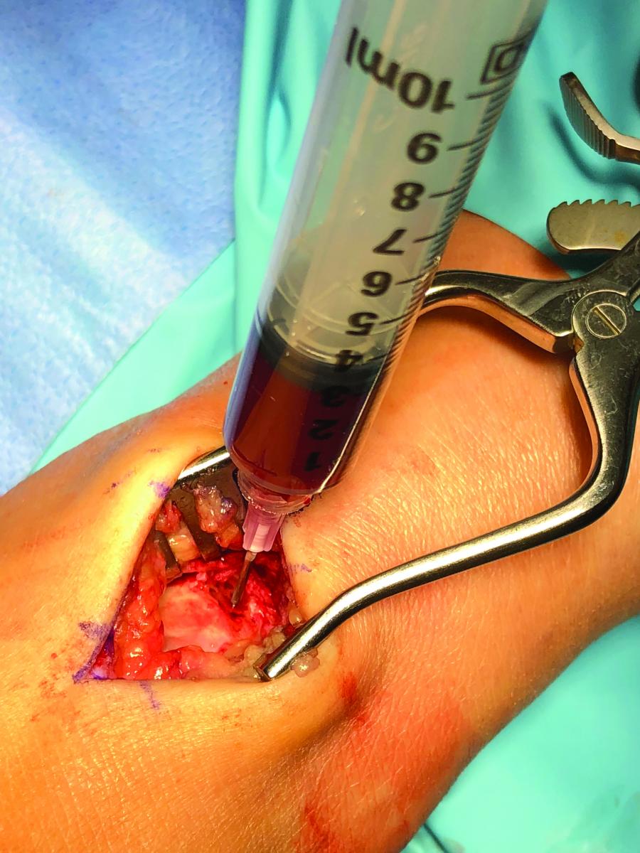
At our institute, we usually inject the subchondral bone. We will at times perform a subchondroplasty with a bone substitute material that hardens and reinforces the region of bone edema. However, we have found bone marrow aspirate injection with microtunnels to work better. Basically, we perform a decompression of the bone and fill the microdecompression with bone marrow concentrate to help with healing. This has been far more successful than subchondroplasty for us. If the cartilage lesion is going to be treated, we perform the subchondral drilling and injection through the lesion, whether it is with a subchondroplasty material or bone marrow aspirate. If the cartilage lesion is minimal and a subchondral region is the main issue with bone edema, we perform the entire procedure from a retrograde approach and do an injection or subchondroplasty retrograde to protect the cartilage.
When it comes to the cartilage and cystic lesion, surgeons most commonly treat this from a anterograde approach through the lesion. However, one could handle the entire process from inside the joint. In rare cases, the cartilage is not damaged and there is only a large lesion. In these cases, a retrograde grafting of the lesion is all that is necessary but this is rare. The majority of large lesions have bone and cartilage involvement.
Emerging Principles With Cartilage Augmentation
Our most common treatment for large lesions with or without bone involvement is cartilage augmentation. Our most preferred material is DeNovo Natural Tissue Graft (Zimmer Biomet). This consists of particles of live cartilage that enable the surgeon to truly lay down the best quality cartilage. However, if we cannot get insurance approval for this material due to cost (about $7,000), we will use the aforementioned BioCartilage product instead.
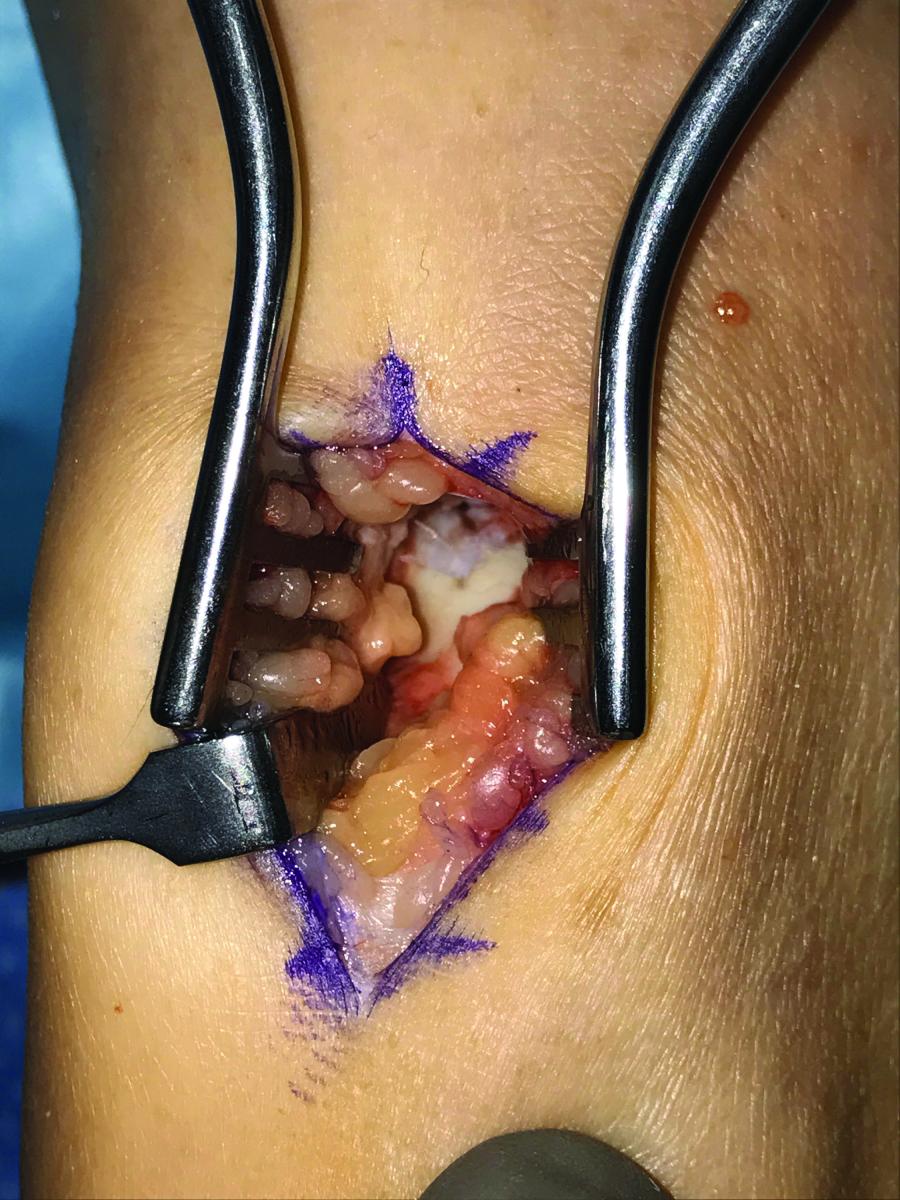
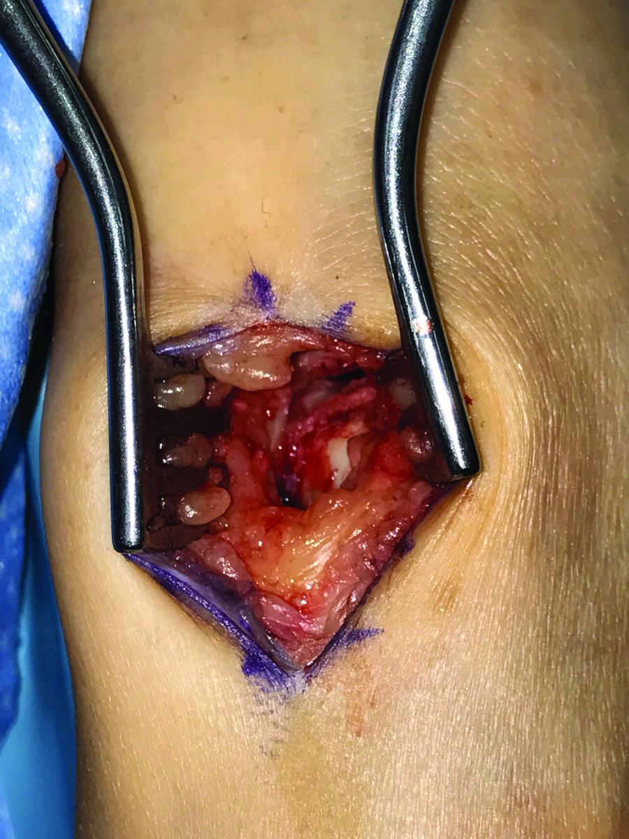
The main difference between DeNovo and BioCartilage is how we deal with the underlying bone. In both cases, I prefer to perform the procedure through a mini-arthrotomy as it is easier to see and treat the lesion and place the cells in the region than it is to do this arthroscopically. In DeNovo cases, we will not damage the underlying subchondral bone and treat the bone lesion or cyst retrograde unless the bone damage involves the subchondral bone, and there is a clear communication with the cystic region. BioCartilage fenestrates and microfractures the subchondral bone, which allows one to fill the underlying bone with a bone graft or bone graft substitute. We prefer taking bone graft with a Jamshidi trocar from the heel and press fitting it into the lesion with the same Jamshidi trocar. We then cover the area with the BioCartilage. If we do a retrograde procedure, we use the same Jamshidi trocar to harvest and then place the bone into a drill hole from the sinus tarsi talus area into the lesion, and fill the lesion.
To summarize, DeNovo is for cartilage lesions and one needs to retrograde drill the cystic lesion needs unless the lesion clearly communicates with the cartilage damage. Surgeons can drill and fill BioCartilage through the ankle joint. If price is not an issue, DeNovo is better than BioCartilage. The main goal with any osteochondral surgery is to deal with any and all issues associated with the talar damage. This would include treating the subchondral bone with drilling and bone marrow aspirate injection, treating the underlying bone lesions with bone great or bone graft substitute, and finally treating the cartilage lesion with a cartilage matrix.
Now that we have dealt with cartilage, what can we do to augment the healing further? I believe some of the best options include an amniotic membrane or some other form of membrane to prevent scarring and augment the release of the hormones necessary for healing.
Another option that we have begun looking at is a dental product called Bioglide (Bioglide). The product is in extensive use in Europe to cover cartilage lesions but in the United States, dentists use it as a cover for bone grafting of teeth prior to implant placement. After a dentist pulls a tooth, he or she uses the Bioglide to cover the area over the bone graft. We use this material off-label over our graft and cartilage regions as a cover. The material stays in place with fibrin glue or one can suture it into place with a couple of stitches. The product provides a happy medium for bone and cartilage growth, and also prevents scar formation, much in the same manner as amniotic membranes.
In Conclusion
Through proper planning and thorough care, one can achieve improved outcomes with osteochondral lesions of the talus. It is important to consider the cartilage, bone and subchondral bone in the complete care of the lesion, and further augment the region with scar prevention mediums. I hope this information will serve as a good jumping off point for further advancement of cartilage repair.
Dr. Baravarian is an Assistant Clinical Professor at the UCLA School of Medicine, and the Director and Fellowship Director at the University Foot and Ankle Institute in Los Angeles (https://www.footankleinstitute.com/podiatrist/dr-bob-baravarian ).
Editor’s note: For further reading, see “Addressing Osteochondral Lesions Of The Talus” in the April 2017 issue of Podiatry Today, “Emerging Concepts In Treating Osteochondral Lesions Of The Talus” in the May 2012 issue or “Orthobiologics: Can They Be Effective For Osteocondral Lesions?” in the June 2007 issue. For other related articles, visit the archives at www.podiatrytoday.com


