Keys To Treating Nail And Digital Trauma
Trauma to the nails and toes can result from various injuries and accidents with complications ranging from onycholysis to amputation. Accordingly, these authors offer pearls on short- and long-term treatment of lower extremity trauma injuries.
Nail and digital injuries can occur from a multitude of traumatic events, including stubbing injuries, crush injuries, puncture wounds, lawnmower accidents and motor vehicle accidents to name a few. The senior author has even treated a toe injury as a result of a patient having his foot run over by an airplane. Digital trauma may result in onycholysis, subungual hematoma, nail bed lacerations, phalangeal fractures or dislocations, soft tissue degloving, traumatic amputation or a combination of the above.
Accordingly, let us take a closer look at some of the presenting pathology when a patient presents to a trauma center or a physician’s office, and discuss keys to treating these patients successfully in order to prevent long-term sequelae.
When Shoe Gear Or Trauma Lead To Onycholysis Or Subungual Hematoma
Some of the most frequently encountered nail trauma may be a result of poorly fitting shoe gear and repeatedly stubbing the nail or other major events as we listed above. Injuries will present as either primary onycholysis or subungual hematoma. Onycholysis, the separation of the nail plate from the nail bed, is also known as a traumatic nail avulsion. The treatment of onycholysis varies depending upon the amount of evulsion that has occurred. One should debride the loose nail back until the nail plate adheres well to the nail bed. In the event the entire nail has been avulsed, remove the nail in total. It is paramount the patient understands a new nail may not appear exactly as the old nail did.
We recommend warm water Epsom salt soaks twice daily with a topical antibiotic ointment and applying a dry dressing after each soak for the first seven to ten days. After the avulsion site and nail bed are well healed, patients can apply a topical antifungal daily or twice daily to prevent mycotic infection as the new nail grows.1 One should also discuss proper nail care and properly fitting shoes with the patient as preventative measures.
A disruption to the vasculature underneath the nail plate may result in the formation of subungual hematoma. The hematoma increases pressure deep to the nail plate and the patient can experience significant pain. It is important to remember that 20 to 25 percent of subungual hematomas are associated with distal phalanx fractures so one should obtain radiographs.1 If the hematoma encompasses less than 25 percent of the nail plate, one may evacuate the blood to alleviate the increased pressure and pain. This can occur by using a handheld cautery unit, an 18-gauge needle, #11 blade or a rotary drill with an appropriate sized bit.1 If the hematoma involves greater than 25 percent of the nail plate, one should avulse the entire nail plate to assess for a nail bed laceration and repair the nail plate appropriately.
Pertinent Insights On Addressing Nail Bed Lacerations
Nail bed lacerations may be simple or complex. Simple lacerations are generally linear or transverse, and complex lacerations may have a stellate appearance. Crush injuries to the digits usually result in more complex lacerations.
The first step in treating nail bed lacerations is to irrigate the laceration thoroughly. One can achieve this with normal saline or a combination of normal saline and antibiotic or antiseptic solution. At our institution, we prefer to combine normal saline with povidone-iodine as a first-line irrigant. Assessing if there is adequate soft tissue coverage and planning your closure prior to suturing will help decrease the chance of complications. For patients who have inadequate soft tissue for repair, surgeons may need to apply a biological dressing to cover the underlying distal phalanx.1
In patients with adequate soft tissue coverage of the distal phalanx, one should attempt primary repair of the laceration. It is important to remember that the nail bed tissue is very fragile and can rip easily if you do not handle it delicately. We recommend repairing nail bed lacerations with absorbable sutures. Removing non-absorbable sutures from a patient’s nail bed can be a very painful experience and the additional trauma to the nail bed may result in additional soft tissue damage. Apply a non-adherent dressing following the repair.
Surgeons should consider any nail bed laceration associated with a distal phalanx fracture as an open fracture and treatment should follow appropriate open fracture protocols. Patients who present to our emergency department with a simple laceration and associated distal phalanx fracture, which is considered a Gustilo-Anderson type 1 fracture, receive a dose of IV antibiotics and usually go home with five to seven days of oral antibiotics in addition to repair of the laceration.2,3 For the phalanx fracture, we immobilize the patient with methods that we will discuss later.
When Patients Lose Soft Tissue In The Foot
Patients may also sustain injuries that result in soft tissue loss of all or part of the digit. Rosenthal developed a classification system for toe tip tissue loss, dividing the digit into different zones.4
Zone 1 is the area of the tissue distal to the distal tip of the distal phalanx. For Zone 1 injuries, one can perform local wound care, use non-adherent dressings and allow patients to heal by secondary intention. Larger defects may require split thickness skin grafting, pinch grafts or biologic dressings.
Zone 2 covers the area of the tissue distal to the nail lunula to the tip of the distal phalanx. These injuries usually require skin flaps for coverage of the bone. Surgeons can do this with V to Y advancement flaps either from the plantar, medial or lateral surfaces depending upon the salvageable soft tissue. The extent of morbidity should always be part of the decision process in determining if a flap will be successful and consideration for a primary distal Symes amputation may be warranted.
 Zone 3 covers the area of the tissue proximal to the lunula. A degloving soft tissue injury in zone 3 is an indication for a primary amputation. Surgeons should amputate back to the level of healthy, viable tissue.
What You Should Know About Digital Fractures
Although most digital fractures are non-displaced, oblique or transverse in nature, surgeons must still employ standard fracture protocols. One should immobilize non-displaced fractures with buddy taping or splinting, and the patient ambulates in an open toe surgical shoe. We allow patients to transition to normal shoe gear as pain tolerance allows. For any phalanx fracture that is displaced greater than 2 mm, angulated or if a joint is dislocated, this requires closed reduction. Closed reduction can usually occur with the patient having local anesthesia.
Following successful reduction and alignment, splint the fractured phalanx as we discussed previously. When treating a hallux fracture, we recommend applying a medial gutter splint made from 2- or 3-inch fiberglass. Due to the amount of force that transmits to the hallux with ambulation, some patients with hallux fractures may require non-weightbearing.
Displaced phalanx fractures that are not amenable to closed reduction require open reduction with fixation. One would determine the type of fixation by the type of injury and fracture pattern. Utilizing AO principles, surgeons can accomplish fixation with K-wires, screws, plates, monofilament wire and/or external fixation.1
Case Study: When There Is A Gunshot Wound With Extensive Soft Tissue Damage
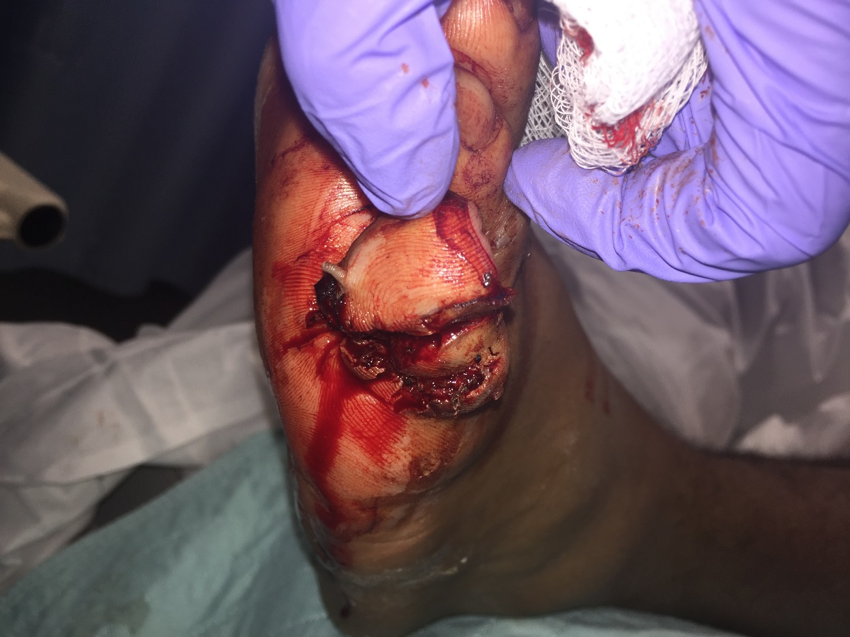
 A 25-year-old male sustained a gunshot wound to his left foot. He had an open plantar wound lateral to the fifth digit with extensive soft tissue damage (see left photo).
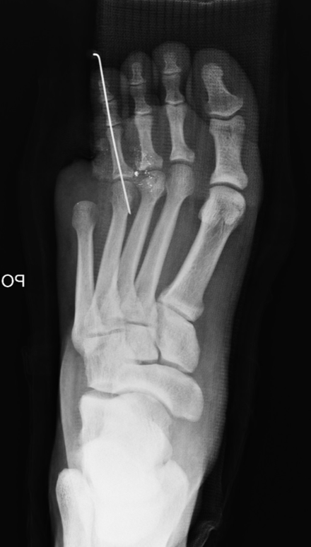
 His pre-op X-rays indicated multiple bullet fragments along with severely comminuted middle and distal phalanx fractures of the fifth toe with bone loss (see right photo). The patient also had a fourth digit dislocation at the metatarsophalangeal joint.
 With consideration of the long-term morbidity associated with the injury, we performed a primary fifth toe amputation (see left photo). We also performed open reduction of the fourth toe dislocation and stabilized the toe with a K-wire. After removing the bullet fragments, we noted a non-displaced third metatarsal head fracture radiographically and addressed this with non-surgical treatment.
Case Study: Treating A Hallux Phalanx Fracture And Dislocated Interphalangeal Joint In An Elderly Patient After A Fall
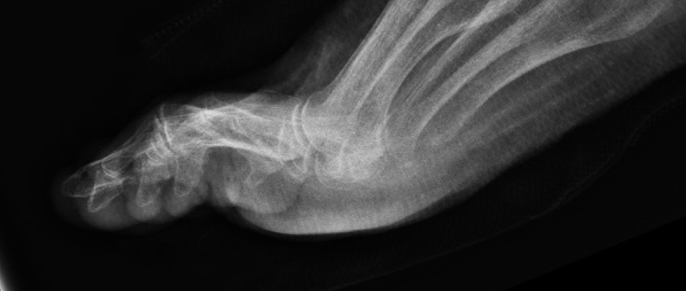
 A 96-year-old female presented following a fall. X-rays show a minimally displaced right hallux proximal phalanx fracture and a plantarly dislocated interphalangeal joint of the hallux (see right photo). She had closed reduction of the dislocation.
 Following closed reduction, we applied a 3-inch fiberglass gutter splint (see left photo). Post-reduction X-rays show a well aligned hallux interphalangeal joint.
Case Study: Repairing A Crush Injury To The Hallux
A patient presented following a crush injury to the left hallux. There is a visible subungual hematoma involving about 50 percent of the nail plate (see right photo).
 Following total nail plate avulsion, we encountered a linear simple laceration of the nail bed. We performed copious irrigation of the laceration and repaired it with absorbable suture material.
Final Considerations
With any injury, the overall health status of the patient must be a consideration. In addition, one must weigh the benefits of heroic reconstruction and the morbidity associated with it versus primary amputation. The surgeon must determine which treatment will have the best long-term outcome for each individual patient, realizing that an amputation may be in the patient’s best interest and an amputation is not always a failure.
As the treating physician, it is also important to convey to any patient with a pedal traumatic injury that the foot or toes may not appear or function the same as they did prior to the injury. Patients must also be aware of any possible sequelae. Sequelae associated with nail and digital trauma includes nail plate malalignment or loss, onychocryptosis, onycholysis, onychomycosis, bacterial infection, splint nail, Beau’s lines, chronic pain and amputation.5
In addition, if a fracture is associated with the injury, malunion or nonunion of a phalanx may occur. The foot and ankle specialist should be familiar with these sequelae and treat nail and digital trauma properly in an effort to decrease the likelihood of complications.
Dr. Cook is the Director of Podiatric Medical Education at University Hospital in Newark, N.J. He is a Fellow of the American College of Foot and Ankle Surgeons.
Dr. Tucker is a second-year resident in the University Hospital Podiatric Medicine and Surgery Residency Program with Reconstructive Rearfoot/Ankle Surgery.
References
- Southerland J. McGlamry’s Comprehensive book of Foot and Ankle Surgery, Fourth Edition. Lippincott Williams & Wilkins, Philadelphia, 2013, pp. 1535–48.
- Gustilo RB, Anderson JT. Prevention of infection in the treatment of one thousand and twenty-five open fractures of long bones: retrospective and prospective analyses. J Bone Joint Surg Am. 1976; 58(4):453–458.
- Gustilo RB, Mendoza RM, Williams DN. Problems in management of type III (severe) open fractures: a new classification of type III open fractures. J Trauma. 1984; 24(8):742–746.
- Rosenthal EA. Treatment of fingertip and nail bed injuries. Orthop Clin North Am. 1983; 14(4):675-697.
- Bharathi RR, Bajantri B. Nail bed injuries and deformities of nail. Ind J Plast Surg. 2011; 44(2):197–202.
For further reading, see the November 2010 DPM Blog “Recognizing And Treating Nail Trauma Caused By Ill-Fitting Shoes” by Tracey Vlahovic, DPM at https://tinyurl.com/l478pak or “Treating Gunshot Wounds In The Lower Extremity” in the January 2014 issue of Podiatry Today.


