Key Pearls On Orthotic Casting And Fabrication
Digital scanning has changed the techniques and outcomes some DPMs use for fabricating orthoses. These panelists explore the advantages to digital versus plaster casting, which biomechanical assessments can maximize casting and how to select the best orthotic material for patients.
Q:
What biomechanical assessments do you perform to optimize casting?
A:
 For Jane Andersen, DPM, the biomechanical exam usually includes assessment of the patient’s resting calcaneal stance position, neutral calcaneal stance position, subtalar joint range of motion and ankle joint dorsiflexion. She also performs a gait exam.
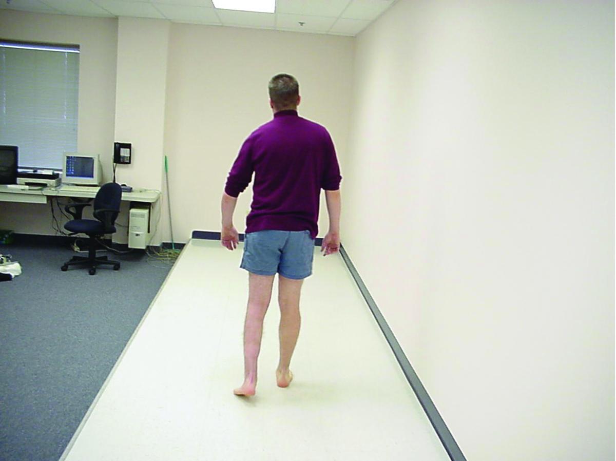
David Levine, DPM, CPed, takes a thorough history regarding all biomechanical issues and complaints from the back down to the foot. In ascertaining the history, he says he is able to key into the biomechanical issues that need his assessment and attention. Dr. Levine conducts a visual inspection for the overall structure of the patient’s thighs, legs and feet, directing his attention to any asymmetric finding. Depending upon the patient and the problem, he will evaluate leg lengths in stance as well as overall posture. Assessing barefoot gait is part of his initial exam as is a range of motion exam of all lower extremity joints focusing on asymmetry.
“It is asymmetry that is often the underlying sign or clue of where problems may emanate,” says Dr. Levine.
Similarly, Roy Mathews, DPM, conducts a physical exam and biomechanical gait analysis, and takes X-rays if necessary. The most common symptoms (such as plantar fasciitis and metatarsalgia) have common causes and he says the assessment of these conditions is not complicated. Dr. Mathews notes it is the uncommon problems that may require F-Scan (Tekscan) gait scanning or video analysis. He likes to tape his patients to simulate orthotic control to determine if taping will help if there are different treatment options.
Q:
Do you prefer scanning or digital casting for orthoses?
A:
Dr. Andersen and Dr. Mathews are currently using digital scanners for their orthoses. Dr. Andersen prefers digital scanning due to the ease of cleanup (or lack thereof). She also says the scanner also speeds up the process in terms of getting the image to the lab.
Dr. Mathews says he can perform a digital scan during his initial exam and use the scan if he determines orthotics are needed following a positive result with taping of the foot. He says the digital scan allows him to capture the image without the patient needing an appointment. He cites the digital method’s efficiency and lack of waste. With a digital scan, he adds that there is no chance of cast deformation during packing or shipping, and it is faster to get the scan to the lab. Digital scans are also environmentally friendly with Dr. Mathews noting they do not require plaster, hot water or shipping of a positive mold.
For Dr. Levine, the preference of digital versus plaster comes from the situation that works most efficiently for the practitioner. “Having my own lab where we are able to control every step from the impressions all the way through the fabrication and dispensing gives us complete control of the entire process,” says Dr. Levine.
Dr. Levine cites advantages to digital scanning, such as evolving improvements in scanning and fabrication technology. He notes digital scans offer faster turnaround times for orthoses, allowing patients to get their devices more quickly.
Q:
What criteria do you use to select the type of material for orthotic fabrication?
A:
Orthotic material selection is dependent upon patients’ footwear, activity level and the biomechanical issues present, according to Dr. Levine. For instance, he notes a hypermobile foot will need more biomechanical control than a foot with a limited range of motion or a range of motion that is more restricted. As Dr. Levine emphasizes, the orthotic material selection can vary depending upon the foot structure and the shoe selection as well. For example, he says a flexible material utilized for a cavus foot will function more rigidly than the same material for a low arched or flat foot. The curvature of the orthotic device decreases the amount of flexibility, according to Dr. Levine.
Similarly, when determining orthotic materials, Dr. Andersen considers factors such as patient conditions, foot type, neurological status, shoe type and activity level/intended activity. Dr. Mathews will also consider the activity the patient is engaged in while wearing the orthosis, which shoe the patient wears for activity or work, and what level of control the patient needs.
In regard to determining the level of control, Dr. Mathews says he prefers to use a thin, composite device that provides 60 percent control and one that the patient can use 90 percent of the time rather than a device that provides 100 percent control but the patient uses only 10 percent of the time.
“I try to manage the control needed and the patient’s desire for shoe gear. It is not always easy,” says Dr. Mathews.
Dr. Andersen is in private practice in Chapel Hill, N.C. She is a Past President of the American Association for Women Podiatrists and is a podiatric expert for Caring.com. She is board-certified in surgery by the American Board of Foot and Ankle Surgery.
Dr. Levine of the Foot and Ankle Specialists of the Mid-Atlantic is in private practice in Frederick, Md. He is also the director and owner of Physician’s Footwear, an accredited pedorthic facility, in Frederick, Md., and the owner of New Balance-Frederick.
Dr. Mathews is in private practice at Vancouver Podiatry in Vancouver, BC. He is affiliated with Vancouver General Hospital and the Royal Columbian Hospital in New Westminster, BC.
Editor’s note: For further reading, see “Point-Counterpoint: Scanner Casting: Is It Better Than Plaster Impression Casting?” in the June 2015 issue of Podiatry Today, “Key Insights On Digital Casting Techniques” in the January 2010 issue or “Insights On The Evolving Nature Of Orthotic Education And Casting” in the June 2014 issue.
To access the archives, visit www.podiatrytoday.com .


