ADVERTISEMENT
A Guide To Cutaneous Manifestations Of Diabetes In The Lower Extremity
Patients with diabetes may be especially susceptible to cutaneous complications including acquired perforating dermatosis, bullous diabeticorum, yellow nails and calciphylaxis. This author provides a guide to diagnosis and treatment of common cutaneous manifestations.
Most, if not all, patients with diabetes will develop skin complications in the course of their disease due to its effect on microcirculation and collagen. The complications of diabetes — including peripheral vascular disease, neuropathy and immune compromise — are implicated in many of the skin conditions associated with the disease.
Despite having common etiologies, though, there are many ways diabetes can manifest in the integument of the lower extremity, from isolated skin lesions to more diffuse rashes to edema and ulceration. Accordingly, let us take a closer look at the various cutaneous manifestations of diabetes as they pertain to podiatric practice, including their etiologies, pathophysiology, prognosis and treatment where possible.
 When Patients With Diabetes Develop Necrobiosis Lipoidica
Necrobiosis lipoidica, also known as necrobiosis lipoidica diabeticorum, is also associated with sarcoidosis, inflammatory bowel disease, autoimmune thyroiditis, rheumatoid arthritis and monoclonal gammopathy. In fact, while 75 percent of patients with necrobiosis lipoidica have or will eventually develop diabetes, only 0.3 to 1.6 percent of patients with diabetes develop this condition.1,2 The etiology is unclear but microangiopathy is the most accepted theory as with many diabetic skin complications.
Necrobiosis lipoidica usually arises between the third and fifth decades of life, and presents as violaceous, red-brown or yellow-brown plaques with an erythematous border, waxy/atrophic surface and prominent telangiectasias on the anterior aspect of the lower legs. Ulceration occurs in 35 percent of patients.2
Due to a lack of quality randomized-controlled trials, there are no evidence-based guidelines for the treatment of necrobiosis lipoidica. Despite the condition’s supposedly strong association with diabetes, tight glucose control has not shown any real benefit in the management of these lesions, thus undermining the condition’s association with diabetes and causing the recently proposed name change to necrobiosis lipoidica.
Steroids are currently the mainstay of treatment. In the case of patients with diabetes, this means intralesional or topical steroids due to concern over glucose control with systemic steroids. While these steroids will control and arrest the inflammatory process, physicians must be careful in their application as they will also further the atrophy that takes place in this condition. This has led to a search for safer yet effective treatments, including pentoxifylline, cyclosporine, aspirin, chloroquine, calcineurin inhibitors, psoralens and ultraviolet light therapy (PUVA), retinoids, hyperbaric oxygen therapy and even skin grafting.1-4 Again, the literature on each is not sufficient to provide solid treatment guidelines but it is promising and the future should bring more effective relief for this condition.
 What You Should Know About Bullous Diabeticorum
Bullous diabeticorum consists of tense, non-inflamed bullae that form abruptly anywhere on the lower extremities. These bullae develop seemingly overnight without trauma and cause little pain or discomfort. The etiology is unknown but microangiopathy is again suspected as a majority of patients have other diabetic complications such as nephropathy and neuropathy. In fact, these bullae may actually be the first sign of underlying glycemic abnormalities. The reported United States incidence is 0.5 percent.1 These diabetic bullae are usually self-limiting and resolve in two to six weeks without scarring as long as there is no secondary infection.
Therefore, treatment is essentially supportive, including compression, aspiration, local wound care and antibiotics if necessary. Many patients never have another episode but future occurrences are not unheard of.2,5
Patients with vitiligo suffer from localized and generalized loss of skin pigment, particularly in the intertriginous areas and orifices of the body. This condition also has an unknown etiology but multiple associations including thyroid disease, Addison’s disease, pernicious anemia, melanoma and autoimmune syndromes. Vitiligo has a reported incidence of 1 percent and affects males and females equally.6 Some studies show that 50 percent of vitiligo cases occur before the age of 20.6 Vitiligo is completely benign and asymptomatic except for its unpleasant cosmetic appearance.
Treatment is difficult. Narrow-band UVB and PUVA are the best treatments, but these modalities require six to 12 months for complete cure even with concurrent topical steroid use.1 Intralesional low-dose steroids showed promise in one small study.6
Key Pointers On Yellow Nails In Patients With Diabetes
Forty to 50 percent of patients with diabetes have yellow nails. Despite their unpleasant appearance, yellow nails are usually not painful or problematic. There is a large list of other associated conditions including lymphedema, infections (pneumonia, AIDS), autoimmune disorders (thyroiditis, rheumatoid arthritis), malignancy (mycosis fungoides, laryngeal carcinoma, gallbladder carcinoma, bronchial carcinoma, breast cancer, non-Hodgkin’s lymphoma, endometrial cancer) and lung disorders such as pleural effusions and bronchiectasis.
Treating the underlying disorder is the key to resolving this condition but half of patients with yellow nails will see spontaneous improvement even without treatment. Patients with resistant cases may need help with vitamin E, 800 IU daily or 5% vitamin E solution drops twice daily.1,2
 Salient Insights On Diabetic Dermopathy
A very common manifestation of diabetes in the lower extremity, diabetic dermopathy develops in 40 percent of patients at some point in the course of their disease. The etiology is again unknown but the theory is that it may be another result of microangiopathy as it frequently arises in conjunction with retinopathy, neuropathy and nephropathy. These lesions begin as round, flat-topped, scaly red papules on the shins that evolve into round, atrophic hyperpigmented lesions of variable size. They are usually asymptomatic but cosmetically unpleasant. Biopsy is not necessary but will show hemosiderin deposition, dermal edema, thickened superficial blood vessels, extravasation of erythrocytes and a mild lymphocytic infiltrate, thus furthering the notion that microangiopathy is the cause.
There is no real treatment available but the lesions may spontaneously resolve.1-2,7
 When Acquired Perforating Dermatosis Arises
Also known as Kyrle’s disease, acquired perforating dermatosis consists of intensely pruritic dome-shaped papules on the lower extremities. It is mostly present in young or middle-aged African-American males.2 Acquired perforating dermatosis is mainly associated with kidney disease and has a prevalence of up to 10 percent.2 However, acquired perforating dermatosis can also arise in patients with diabetes, heart disease or hepatitis, or it can be idiopathic. These lesions have central keratotic plugs that will show up on biopsy as keratin and necrotic debris.
Controlling the systemic disease will certainly help but patients with resistant cases may also need topical steroids and an antihistamine. There is limited evidence supporting the use of allopurinol 100-300 mg three times daily.8 Other supplemental treatments include retinoids, vitamin A, UV light therapy, topical keratolytics and cryotherapy.2,8-10 Additionally, one should inform patients that while the lesions are chronic, they can heal if patients avoid trauma and scratching.
 What You Should Know About Calciphylaxis
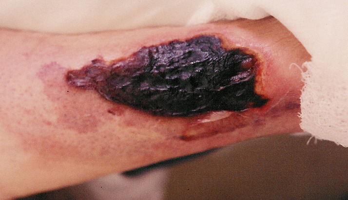
Calciphylaxis is an ischemic small-vessel vasculopathy that is also known as calcific uremic arteriolopathy. It mostly arises in hemodialysis patients (1 to 4 percent prevalence) but also occurs in patients with diabetes, primary hyperparathyroidism, alcoholic liver disease, malignancy, and protein C&S deficiencies. Patients with diabetes in renal failure are at particular risk.
Calciphylaxis manifests as extremely painful necrotic ulcerations on the feet and legs. While biopsy is not necessary for the diagnosis, it will show cutaneous necrosis with fibrin thrombi, endovascular fibrosis and calcification.
Treatment consists of 25 g of intravenous sodium thiosulfate after each dialysis session along with pain control. The majority of patients with calciphylaxis will have temporary improvement but only 25 to 30 percent of calciphylaxis ulcerations will completely resolve. Sadly, even with treatment, the one-year mortality rate is between 45 and 80 percent.11
Pertinent Pearls On Treating Eruptive Xanthomas
Eruptive xanthomas occur in less than 0.1 percent of patients with diabetes.2 As their name suggests, these lesions form yellow lipid deposits in the skin and tendons, mostly on extensor surfaces and the Achilles tendon. They may have an erythematous halo as well. Eruptive xanthomas are not usually painful unless they are prominent and cause pressure. Besides diabetes, these lesions are also associated with dyslipidemias, hypothyroidism, Cushing’s disease, alcoholism, systemic lupus erythematosus, renal disease, liver conditions, and certain medications such as beta-blockers and estrogen medications.
Skin xanthomas mostly resolve on their own once there is control of lipid levels but tendinous xanthomas tend to persist and may require surgical excision.1
When Patients With Diabetes Have Lichen Planus
Lichen planus consists of violaceous hyperpigmented papules primarily affecting the ankles and wrists. The etiology is unknown but there are multiple variants including a palmoplantar version that is less common and more difficult to treat. It can even affect the nails, causing thinning, splitting and pterygium formation until the entire nail is destroyed and scarred. Pruritus is often severe. Many cases are self-limiting but resistant cases may require topical or even systemic steroids. In the case of nail involvement, injections of triamcinolone (Kenalog, Bristol-Myers Squibb) into the nail matrix may be necessary.10
Palmoplantar lichen planus is a much less common variant that does not resemble classic lichen planus in that it does not demonstrate Wickham’s striae and the typical polygonal lesion. As its name implies, palmoplantar lichen planus is mostly localized to the soles and palms. Treatment is more difficult and large randomized controlled trials are almost non-existent. Physicians have attempted treatment with various modalities, including retinoids, enoxaparin, steroids, calcineurin inhibitors, cyclosporine and surgery. Small case studies have shown efficacy with the aforementioned treatments but none have proven universally successful in larger studies.12
Why You Should Be Wary Of Diabetic Foot Ulcerations
No discussion of the cutaneous manifestations of diabetes is complete without talk of diabetic ulcerations. Patients with diabetes have an almost 20 percent lifetime risk of developing foot and leg ulcers.10 Ulcerations in a patient with diabetes are significant because of the high risk of infection and amputation. Indeed, over 80 percent of non-traumatic amputations are attributed to diabetic ulcerations. There are various types, including but not limited to decubitus, venous, arterial and calciphylaxis ulcerations as I described above.10
A wise podiatrist once said, “Treating wounds is 95 percent etiology, 5 percent product.”13 Accordingly, treatment involves correcting the pathophysiology that caused the wound in the first place. When it comes to decubitus wounds, which are usually in high-pressure areas such as the forefoot and have significant callus surrounding them, there should be appropriate offloading via padding, shoe inserts, braces, boots, casts or even a period of non-weightbearing if necessary.
Refer patients with arterial wounds for angioplasty and possible stenting to increase the arterial flow, and provide improved nutrition for the affected wound. Venous wounds need compression to relieve the lower extremity of the excess fluid that causes tightness of the tissue and predisposes to traumatic ulcerations. Treat infections with antimicrobials as necessary and excise malignancies where possible.
Key Pointers On Cutaneous Infections
We all know the statistics when it comes to diabetic wound infections but 20 to 50 percent of patients with diabetes will develop other types of cutaneous infections as well. The same comorbidities (such as peripheral vascular disease, neuropathy and immune compromise) that put patients at risk for wound infections also predispose them to other cutaneous infections. Common infections include Candida, dermatophyte onychomycosis/tinea pedis, impetigo, folliculitis, carbuncles, furunculosis, ecthyma, erysipelas and erythrasma.
Oral antimicrobials may be necessary in severe cases but frequently, topical antibiotics or antifungals are effective.2
When There Are Cutaneous Side Effects With Diabetes Medications
Sometimes the very medications used to treat diabetes can have adverse effects on the integument. While the side effects may be temporary, some cases may require discontinuation of the offending drug and consideration of an alternative modality.
For example, metformin can have effects on the dermis such as psoriasiform drug eruptions, erythema exsudativum multiforme, leukocytoclastic vasculitis, photosensitivity, erythema, exanthema, pruritus and urticaria. Rosiglitazone (Avandia, GlaxoSmithKline) and pioglitazone (Actos, Takeda Pharmaceuticals), on the other hand, can cause edema of the lower extremities. The DPP-4 inhibitors such as sitagliptin (Januvia, Merck) can cause hives/rash and facial swelling while sulfonylureas like chlorpropamide, tolbutamide and glimepiride (Amaryl, Sanofi) also can cause skin rash. The SGLT-2 inhibitors such as empagliflozin (Jardiance, Boehringer Ingelheim) and canagliflozin (Invokana, Janssen Pharmaceuticals) can cause yeast infections of the groin. One should work up any new rash or swelling in a patient with diabetes accordingly, and strongly consider medication side effects.2,14
In Conclusion
Diabetes has many effects on the body and the skin is no exception. There are plenty of cutaneous manifestations that can occur due to microangiopathy, neuropathy and immunocompromise as well as other complications. Frequently, blood sugar control can help but sometimes topical or systemic medications such as steroids are necessary. In a few cases of cutaneous manifestations, no treatment is universally successful and the patient must simply tolerate the condition. Either way, the proper diagnosis of these cutaneous manifestations is essential in order to determine prognosis and treatment options, and provide for good outcomes and the patient’s peace of mind.
Dr. Vella is in private practice in Gilbert, Ariz.
References
- Habif TP. Cutaneous Manifestations of Internal Disease. In: Clinical Dermatology: A Color Guide to Diagnosis and Therapy. Fifth Edition. Mosby, Elsevier, China, 2010, pp. 974-998.
- Van Hattem S, Bootsma AH, Thio HB. Skin manifestations of diabetes. Cleve Clin J Med. 2008; 75(11):772-87.
- Feily A, Mehraban S. Treatment modalities of necrobiosis lipoidica: a concise systematic review. Dermatol Reports. 2015; 7(2):5749.
- Reid SD, Ladizinski B, Lee K, et al. Update on necrobiosis lipoidica: a review of etiology, diagnosis, and treatment options. J Am Acad Dermatol. 2013; 69(5):783-91.
- Bhutani R, Walton S. Diabetic bullae. Br J Diabetes Vasc Dis. 2015; 15: 8-10.
- Wang E, Koo J, Levy E. Intralesional corticosteroid injections for vitiligo: A new therapeutic option. J Am Acad Dermatol. 2014; 71(2):391-3.
- Finucane KA, Archer CB. Recent advances in diabetology: diabetic dermopathy, autoantibodies in the prediction of the development of type 1 diabetes, and islet cell transplantation and inhaled insulin as treatment for diabetes. Clin Exp Dermatol. 2006; 31(6):837-40.
- Shih C, Tsai T, Huang H, et al. Kyrle’s disease successfully treated with allopurinol. Int J Dermatol. 2011; 50: 1170-1172.
- Saray Y, Seckin D, Bilezikci B. Acquired perforating dermatosis: clinicopathological features in twenty-two cases. J Eur Acad Dermatol Venereol. 2006; 20:679-688.
- Vlahovic T, Schleicher SM. Skin Disease of the Lower Extremities: A Photographic Guide. HMP Communications, Malvern, PA, 2012.
- Nigwekar SU, Brunelli SM, Meade D, et al. E. Sodium thiosulfate therapy for calcific uremic arteriolopathy. Clin J Am Soc Nephrol. 2013; 8(7):1162-1170.
- Feily A, Yaghoobi R, Nilforoushzadeh MA. Treatment modalities of palmoplantar lichen planus: a brief review. Adv Dermatol Allergol. 2016; 33(6):411-415.
- Personal conversation with Brent Bernstein, DPM, Aug 2014.
- Omudhome O. Type 2 oral diabetes medications (oral). Medicine.net. Accessed Jan. 4, 2017.


