Essential Considerations With Heel Ulcers And Diabetic Limb Salvage
Rearfoot ulceration is a pinnacle of complexity in diabetic limb salvage. It is a perverse pathology reserved for those with the most severe systemic illnesses. An unfortunate outcome of a merciless disease, heel ulceration afflicts our most brittle patients. It debilitates them while systemic manifestations work their way from accessory limitations to end-stage organ failure. Multifaceted barriers to intervention often build upon each other to block healing. Stopping the process before it robs patients of life and limb is crucial, but has challenged surgeons for decades.
The risk for major lower extremity amputation in patients with diabetic foot ulcers (DFUs) is significant and even worse for those who have a DFU in the heel.1-4 The heel, specifically, with its proximity to critical kinematic structures, desperately jeopardizes these mechanisms and threatens almost all attempts at offloading. There is elevated complexity when tissue depletion finally permits the extension of osteomyelitis to the calcaneus.
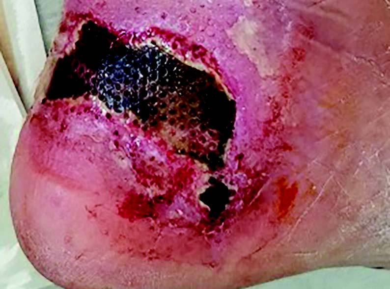
These are maximally threatening cases with a 52 percent chance of major lower extremity amputation.5 For those with sustained immobility who develop decubitus-type ulcers, this translates to an almost twofold higher mortality rate in comparison to those without heel ulceration (see first photo above).6
For good reason, evolution provides a unique morphology of the heel to protect against this outcome. The thick stratum corneum and elastically bound adipocytes, well known to foot surgeons, provide protection but at a metabolic cost.7 Fed by dually redundant macro- and highly anastomotic microvascular supplies, tissue in healthy hosts provides a lifelong barrier despite the grueling cyclic loads.8 Failure begins when expansive comorbidity profiles deplete perfusion and glycosylate neural feedback loops. Shear and biomechanical aberrations contribute, and tissue breakdown ultimately prevails.
In patients who develop decubitus-type ulceration, the level at which breakdown occurs is less understood. Despite focal pressure loading at the weightbearing prominence of the calcaneus, the precise depth at which breakdown occurs is likely more superficial. The panniculus carnosus, a thin, striated muscular layer which has evaded evolutionary revision, is highly suspect in this extended compression.8 Avascular myolysis of this one mm thick structure becomes a nutrient-rich, safe harbor for bacterial colonization and necrosis that extends from there.
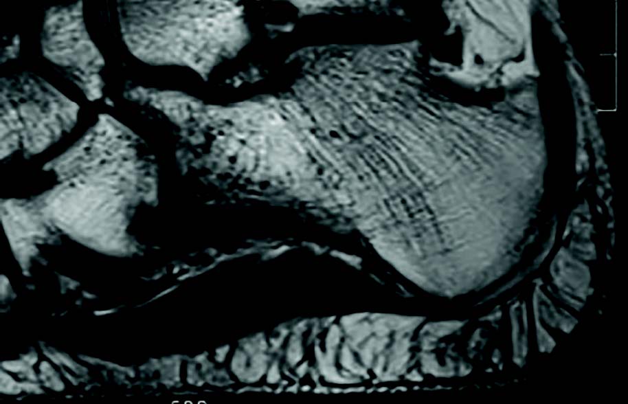
The plantar fat pad does not help in either case. The elliptical adipocytes, perpendicular to the weightbearing surface of the heel and tightly constrained by fibrous septae, are largely avascular (see second image above). This enhances failure in two ways. First, the fibrous channels permit longitudinal bacterial communication. Transmission from deep to superficial tissue planes and vice versa allows rapid tissue degradation, resulting in full-thickness liquefaction. Second, as local trauma rages, the tightly loculated adipocytes swell. In a cascade of cytokines and interleukins, perfusion to this already avascular structure slows.7 Eventual edema stymies local perfusion pressure and a microscale compartment syndrome ensues. Gross necrosis and tissue loss prevail.
Critical as it is, soft tissue envelope loss merely is an erosion of this bacterial barrier. The grave severity comes when deep infiltration permits environmental extension to the osseous edge. The bacterial trek permeates the cortex, osteomyelitis ensues and the complexities of successful salvage drastically expand.
Recognizing Key Diagnostic And Treatment Challenges With Heel DFUs
An early diagnosis for osteomyelitis is essential but pathologic complexities continue to ravage attempts at success. Recent work by Meyr and coworkers questions the nominal results of the Jamshidi needle bone biopsy.9 Specimen sampling and lab processing discrepancies plainly exist. Consistent guidelines for handling remain elusive.
There is diagnostic ambiguity as well. In the groundbreaking 2011 study, Meyr and colleagues surveyed four board-certified pathologists and assessed their histologic analysis of bone biopsy specimens.9 They found there was only agreement between the diagnosticians for a third of the samples. This is shocking to most surgeons. A new method brings an evidence-based approach to gut instinct. The International Working Group on the Diabetic Foot recommends stratifying the definitive diagnosis of osteomyelitis by likelihood and calls on multiple objective inputs such as imaging, culture and pathology reports for collaborative confirmation.10
Outside the local and systemic septic effects of ulceration, physical deconditioning can be a second hit, especially if long-term attempted intervention remains stagnant. Prescribed functional limitations are easy to extend when ulcer repair is evident but can be problematic if progress stalls. Recovery is possible in certain populations. These include young, active individuals whose systemic diseases are well-controlled. For others, especially those with systemic compromise prior to ulceration, rehabilitation potential reaches a critical point after continued ambulatory weakening. Recovery to pre-ulcer function, even after achieving successful salvage, may not be possible.11
The unfortunate reality of failed intervention, both major lower extremity amputation and non-function, is mortality, which is well above the baseline in the diabetic population.12,13 Reports of increasing cause-specific mortality in those with pedal ulceration continue to emerge. Non-ambulation is a strong predictor of mortality in this group.14-16 The risk is even higher for those who undergo terminal amputation. In a 2007 meta-analysis, Stern and coworkers found that non-ambulation increased mortality at least twofold following major lower extremity amputation.17
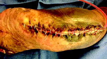
There is hope as there are several procedures have emerged to help modify these risks. Novel techniques such as the vertical contour calcanectomy have emerged as tertiary limb-sparing interventions with initial success.18 Aggressive resection provides an avenue for salvage even after multi-intervention failure suggests terminal amputation to be the likely outcome. Massive volume reduction of the calcaneus can reduce osseous bioburden and accommodate focal envelope loss. Large incisions with wide margins allow thorough debridement, ulcer excision and, in some cases, primary closure (see third photo above).
Final Thoughts
Despite assertive procedures like this, heel ulceration remains high risk. Rapid and accurate diagnosis, aggressive intervention and implementation of novel surgicaltechniques are essential facets for success.An emphasis on reestablishing mobility is guiding further innovation. Until the development of technology that can detect, accommodate and prevent tissue breakdown, aggressive surgical management of heel ulceration will be the mainstay of successful salvage.
Dr. Kennedy is a current Limb Salvage Fellowat MedStar Georgetown University Hospital in Washington, D.C.
1. Pecoraro RE, Reiber GE, Burgess EM. Pathways to diabetic limb amputation. Basis for prevention. Diabetes Care. 1990;13(5):513–521.
2. Apelqvist JLJ, Agardh CD. Long-term prognosis for diabetic patients with foot ulcers. J Int Med. 1993;33:485-491.
3. Carsten C, Taylor S, Langan E, Crane M. Factors associated with limb loss despite a patent infrainguinal bypass graft. Am Surg.1998;64(1):33-38.
4. Younes N, Albsoul A, Awad H. Diabetic heel ulcers: a major risk factor for lower extremity amputation. Ostomy Wound Manage. 2004;50(6):50-60.
5. Faglia E, Clerici G, Caminiti M, Curci V, Somalvico F. Influence of osteomyelitis location in the foot of diabetic patients with transtibial amputation. Foot Ankle Int. 2013;34(2):222–227.
6. Lyder CH AE. Pressure ulcers: a patient safety issue. In: Hughes RG, ed. Patient Safety and Quality: An Evidence-Based Handbook for Nurses. Rockville, MD:Agency for Healthcare Research and Quality;2008.
7. Arao H, Shimada T, Hagisawa S, Ferguson-Pell M. Morphological characteristics of the human skin over posterior aspect of heel in the context of pressure ulcer development. J Tissue Viability. 2013;22(2):42-51.
8. Cichowitz A, Pan WR, Ashton M. The heel: anatomy, blood supply, and the pathophysiology of pressure ulcers. Ann Plast Surg. 2009;62(4):423-429.
9. Meyr AJ, Singh S, Zhang X, Sheridan MJ, Khurana JS. Statistical reliability of bone biopsy for the diagnosis of diabetic foot osteomyelitis. J Foot Ankle Surg. 2011;50(6):663-667.
10. Lipsky BA, Aragon-Sanchez J, Diggle M, et al. IWGDF guidance on the diagnosis and management of foot infections in persons with diabetes. Diabetes Metab Res Rev. 2016;32(Suppl 1):45-74.
11. Attinger CE, Brown BJ. Amputation and ambulation in diabetic patients: function is the goal. Diabetes Metab Res Rev. 2012;2(Suppl 1):93–96.
12. Silbernagel G, Rosinger S, Grammer TB, et al. Duration of type 2 diabetes strongly predicts all-cause and cardiovascular mortality in people referred for coronary angiography. Atherosclerosis. 2012;221(2):551-557.
13. Seshasai SRK, Kaptoge S, Thompson A, et al. Diabetes mellitus, fasting glucose, and risk of cause-specific death. N Engl J Med. 2011;364(9):829–841.
14. Saluja S, Anderson SG, Hambleton I, et al. Foot ulceration and its association with mortality in diabetes mellitus: a meta-analysis. Diabetic Med. 2020;37(2):211–218.
15. Wannamethee SG, Shaper AG, Whincup PH, Lennon L, Sattar N. Impact of diabetes on cardiovascular disease risk and all-cause mortalityin older men: influence of age at onset, diabetes duration, and established and novel risk factors. Arch Intern Med. 2011;171(5):404–410.
16. Brownrig JRW, Davey J, Holt PJ, et al. The association of ulceration of the foot with cardiovascular and all-cause mortality in patients with diabetes: a meta-analysis. Diabetologia. 2012;55(11):2906–2912.
17. Stern JR, Wong CK, Yerovinkina M, et al. A meta-analysis of long-term mortality and associated risk factors following lower extremity amputation. Ann Vasc Surg. 2017;42:322–327.
18. Elmarsafi T, Pierre AJ, Wang K, et al. The vertical contour calcanectomy: an alternative surgical technique to the conventional partial calcanectomy. J Foot Ankle Surg. 2019;58(2):381-386.


