Emerging Insights On First Ray Hypermobility
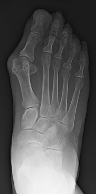
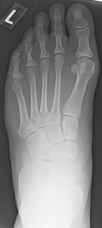
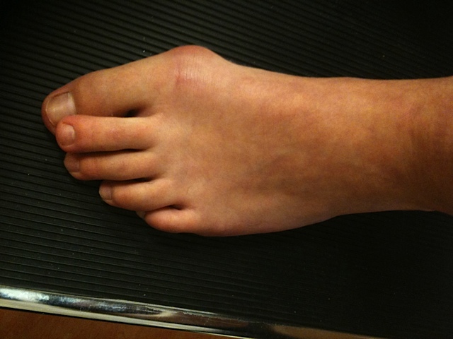
 Hypermobility of the first ray is a critical component in addressing hallux valgus. Accordingly, these authors examine the research on assessing hypermobility and discuss pertinent considerations in achieving optimal surgical correction while preventing recurrence.
There has been wide ranging discussion on the effect of hypermobility in the development of hallux valgus, what we consider normal versus pathologic and the role this plays in determining the appropriate treatment. At the foundation, there is controversy over how to measure hypermobility in both static and dynamic states, and in what plane. A previous review of the literature brings to light the difficulties of assessing and treating hypermobility, and the wide range of theories.1
Accordingly, we would like to review current concepts regarding hypermobility and the importance of this factor in the correction of a hallux abducto valgus (HAV) deformity. We will also discuss the significance of rotation of the first metatarsal in the frontal plane and how this may be altering our perception of where the abnormal motion is actually occurring.
The role of the first ray in gait is to create a stable support for propulsion so the body is able to move forward effectively.2 Root and colleagues described hypermobility of the first ray as an abnormal dorsiflexion motion in response to ground reactive forces. Although the vast majority of pathologic motion is usually in the sagittal plane, Root correctly described the motion as triplane motion with the metatarsal rotating in a valgus direction. Root and colleagues believed that hypermobility was either congenital or acquired, and that acquired hypermobility was a result of pronatory forces of the foot.3 To reinstate a stable construct for propulsion and concurrently treat the hallux abducto valgus (HAV) deformity, a tarsometatarsal joint fusion may be indicated when hypermobility of the first ray is visible upon evaluation of a patient.4
How Our Understanding Of Hypermobility Has Evolved
There has been a tremendous amount of research dedicated to studying hypermobility. Roukis and coworkers reviewed 70 papers in 2003 and did not find a consensus regarding the measurement of normal or abnormal first ray range of motion, planes of mobility, or techniques of measurement.1
Since 2003, our understanding of hypermobility has led to the conclusion that other bones besides the first metatarsal and medial cuneiform may contribute to this condition. Fusion is frequently necessary to control excessive motion but can also compensate for malpositioning of the first metatarsal. In other words, simply locking the metatarsal cuneiform joint may not be sufficient to correct the excess motion and eliminate the deformity. Hypermobility throughout the Lisfranc joint, ineffective dynamic locking of the entire medial column and inadequate tendon and ligament integrity may contribute greatly to collapse of the arch and excessive motion, particularly in stance and propulsive phases of ambulation.
A study by Kamanii and colleagues looked at 20 people who were diagnosed with joint hypermobility syndrome according to Beighton and Bulbena score models.5,6 Comparing these people to 20 healthy controls, the researchers obtained weightbearing anteriorposterior (AP) and lateral radiographs, and assessed 11 angles. The authors found a statistically significant difference in calcaneal pitch and tarsometatarsal angles on lateral radiographs, and the first metatarsophalangeal joint (MPJ) angles on AP radiographs. From their data, the study authors suggested that people with hypermobility were predisposed to having a pes planus foot type and HAV deformities.
 King and Toolan compared 15 healthy people without HAV to 25 individuals who failed conservative treatment for HAV.7 The investigators compared clinical evaluations between the two groups for hypermobility. They determined hypermobility using two methods. They assessed dorsiflexion of the first ray relative to the lesser rays with manual dorsiflexion force and subsequently assessed overloading of the second ray as determined by a plantar hyperkeratotic lesion. Researchers evaluated weightbearing AP and lateral radiographs for both groups to compare the distal metatarsal articular angle, intermetatarsal angle, MPJ angle, talonavicular coverage and the talo-first metatarsal angle. On the lateral radiographs, the study authors assessed the lateral MPJ angle, the talo-first metatarsal angle and Meary’s angle.
The authors also evaluated two new parameters for measuring hypermobility on the lateral radiographs.7 The first was the perpendicular distance between the plantar aspect of the medial cuneiform and the plantar aspect of the first metatarsal. The second technique was to measure the angle formed by the medial cuneiform and first metatarsal articular surface, extending from the superior to the inferior aspect of the joint. All the patients with HAV had clinical signs of hypermobility. In comparing patients with HAV to healthy people, the study findings revealed statistically significant differences in the distal metatarsal articular angle, the intermetatarsal angle, the MPJ angle, talonavicular coverage, the lateral talo-first metatarsal angle, the dorsal translation of the first metatarsal, the medial cuneiform/first metatarsal articular angle, and the lateral MPJ angle.
King and Toolan concluded that there was a correlation between radiographic and clinical testing for hypermobility as well as an indication for lateral imaging in determining hypermobility.7 They suggest that measurement of the dorsal translation of the first metatarsal and the medial cuneiform/first metatarsal articular angle could represent “quantifiable radiographic measures” for defining hypermobility.
Glasoe and coworkers compared two devices that both measure first ray motion in the sagittal plane.8 Both are similar in that they apply a force against the plantar first metatarsal and measure the dorsal translation from a probe at the dorsal aspect of the first metatarsal. The Klaue device applies a manual force and the Glasoe device applies a quantifiable force using a screw mechanism. In this study involving 40 at-risk patients, the authors found that the average dorsal translation was 5.2 (±1.5) mm with the Klaue device and 4.9 (±1.6) mm with the Glasoe device. The authors did not find a statistically significant difference in measurements between the two devices and determined each device was able to detect significance within 1 mm.
The study authors concluded the Klaue device was appropriate for clinical investigators while the Glasoe device was better for studies requiring specific force measurements.8 The researchers also concluded that 8 mm of motion may be a threshold for hypermobility as it is two standard deviations greater than the at-risk population they examined.
 In a study assessing methods of measuring hypermobility, Kim and colleagues compared the Eulji Medical Center (EMC) device to two well-known alternative methods.9 The EMC device consists of two separate “L” shaped objects with the first being a measuring device held on the dorsal aspect of the second ray and the second component held over the first ray. The first ray component lined up next to the measurement guide component on the second ray so researchers could measure the movement in millimeters before and after manual dorsiflexion of the first ray. The study authors compared the EMC device to the Coleman block method in 69 feet and the Klaue device in 46 feet. There were significant differences in measurements between the EMC device and Coleman block test, but the authors noted a correlation coefficient of 0.84. The differences in measurements between the EMC device and Klaue device were not significant, and showed a correlation coefficient of 0.924. Kim and coworkers concluded that the EMC device provided a valid result and could easily assess hypermobility.
Singh and colleagues evaluated 600 feet divided into one group of 187 feet with a HAV deformity and one group of 413 control feet.10 On every foot, researchers measured the first ray range of motion using the Klaue device. They altered the device to not only obtain dorsal mobility but dorsal medial mobility as well at a 45-degree angle. The results showed that mobility in the 45-degree plane was significantly higher than in the dorsal plane for the control group. In the HAV group, 57.8 percent of the patients were hypermobile in both the dorsal and 45-degree angular plane. Another 23.5 percent of patients in the study were only hypermobile in the 45-degree angle plane. The authors concluded that evaluation of dorsal medial plane mobility along with dorsal mobility is preferable to only measuring dorsal plane mobility when evaluating for hypermobility.
Key Pointers On Managing Hypermobility
Each of the aforementioned studies has demonstrated that there is a wide range of variations in the quantity and plane of motion associated with hypermobility. In the presence of hypermobility, the correction of HAV generally depends on blocking motion of the first metatarsal by holding it to a single position while the rest of the foot retains some degree of motion. Consequently, there are many options for the correction of HAV. However, choosing the appropriate procedure for a specific patient is critical to achieve the desired correction while preventing recurrence.
Coetzee and coworkers studied 24 patients (26 bunions) who had recurrence of their bunion deformity after distal, shaft and proximal osteotomies.11 They found that nine patients had severe hypermobility and 12 had mild to moderate hypermobility. The authors found a disproportionate group of recurrences among those patients with clinically diagnosed hypermobility. They concluded that hypermobility may be related to a higher rate of bunion recurrence when surgeons do not address it with an arthrodesis of the first tarsometatarsal joint.
Pentikainen and coworkers looked at 100 patients 7.9 years after a distal osteotomy procedure.12 While 23 patients were lost to the final follow-up, the study authors found a recurrence of deformity, which they described as a hallux valgus angle of >15 degrees, in 73 percent of the remaining patients.
However, in another study, Coughlin and colleagues found a statistically significant reduction in first ray mobility after a proximal crescentic osteotomy and distal soft tissue reconstruction in 103 patients (122 feet) who had moderate or severe primary hallux valgus.13 They noted an improvement of pain on the Visual Analog Scale from a preoperative value of 6.5 to a two-year follow-up postoperative value of 1.1. They also noted an improvement in the American Orthopedic Foot and Ankle (AOFAS) score from 57 preoperatively to 91 postoperatively.
 In their assessment of cadaver models, Rush and colleagues found that when a HAV deformity was present, dorsal drift was present with applied force due to the inability of the windlass mechanism to activate.14 They noted that a proximal osteotomy reduced this dorsal migration by 26 percent with realignment and restoration of the functional stability. They concluded that hypermobility was a symptom of the HAV deformity and one can correct it in a cadaver model without an arthrodesis of the first tarsometatarsal joint.
From the high rate of recurrence, we can see that correction of HAV in the presence of hypermobility is not as simple as constraining the first metatarsal in space. In fact, the current thinking about the HAV deformity may not consider hypermobility as an important indicator for treatment. Dayton and colleagues state that osteotomies are mainly treating only the reduction of the intermetatarsal angle.15 They suggest that the deformity correction should take place at the center of rotation of angulation (CORA) as described by Paley and coworkers.15,16 The area of correction then would exist at the metatarsal cuneiform joint and thereby not create a secondary CORA with an osteotomy.15 Dayton and colleagues also address the frontal plane or “third plane” of the bunion deformity by derotating the first metatarsal in the frontal plane with the first tarsometatarsal arthrodesis.17 The frontal plane of deformity is visible on the sesamoidal axial radiographs as a valgus rotation of the metatarsal.
Most recently, valgus rotation of the metatarsal has become a critical element in our understanding of the cause of HAV and the role of hypermobility. Plain radiographs that show an elevated tibial sesamoid position imply that the metatarsal has migrated laterally, resulting in a bowstringing effect of the flexor tendon. However, if one considers the sesamoid position on a sesamoid axial view, it is apparent that the sesamoid remains within the sesamoid grooves in contact with the metatarsal head. Instead, the entire metatarsal rotates into a valgus position.
This brings an entirely new perspective to the term hypermobility. The rotation of the metatarsal itself gives the impression of lateral deviation of the sesamoids on the AP radiograph.15 Therefore, with triplanar correction of the bunion deformity at the metatarsal cuneiform joint, one can achieve the necessary correction at the center of rotation of angulation. By addressing the valgus rotation of the metatarsal in addition to the increased metatarsal angle and the decreased metatarsal declination angle, the metatarsal head appears to move into a more correct position. In many cases, this can eliminate the need for additional soft tissue balancing such as adductor hallucis tendon releases and lateral collateral fibular sesamoid ligament releases.15,17
The concept of metatarsal rotation raises some questions about the significance of sagittal and transverse plane hypermobility. In order to adequately correct the intermetatarsal angle and return the sesamoids to a functional position, Dayton and colleagues suggest that derotation of the metatarsal with fusion of the joint is necessary, and would inherently correct the hypermobile joint.17
In Conclusion
Hypermobility is a much more complex influence on the development of HAV than we previously thought. In some cases, there is clearly a simple collapse of the first metatarsal resulting in intermetatarsal splaying and medial arch collapse. However, in other cases, one can appreciate that triplane motion dominates and simple stabilization of the first metatarsal-medial cuneiform joint may be insufficient. The high rate of HAV recurrence after surgery and the complexity of the types of motion that can occur at the first metatarsal in both stance and ambulation suggest that other forces are in play to cause HAV. Based on these observations, it is clear that a more comprehensive pre-surgical assessment is probably needed to predict where the excessive motion and lack of stability is coming from.
As we gain a better understanding of the role of varus/valgus rotation of the first metatarsal, we will become better at predicting when this motion should be limited as well. If one can accurately determine the CORA, then a procedure such as a modified Lapidus procedure will potentially control the sagittal plane hypermobility while derotating the metatarsal to counter that type of malpositioning as well. As surgeons gain a better understanding of the interactions between the sesamoid apparatus and the first metatarsal, it is likely that the techniques used to manage hypermobility will evolve as well.
Dr. Bohman is the Chief Podiatric Surgical Resident and an Instructor in Surgery at Harvard Medical School. She is also affiliated with Cambridge Health Alliance in Cambridge, Mass.
Dr. Landsman is the Chief of the Division of Podiatric Surgery and an Assistant Professor of Surgery at Harvard Medical School. He is also affiliated with Cambridge Health Alliance in Cambridge, Mass.
References
- Roukis TS, Landsman AS. Hypermobility of the first ray: a critical review of the literature. J Foot Ankle Surg. 2003;42(6):377-90.
- Valmassy RL. Clinical Biomechanics of the Lower Extremities. Mosby, St. Louis, Missouri, 1996, pp. 22-23.
- Root ML, Orien WP, Weed JH. Motion of the joints of the foot: the first ray. In Root SA (ed.) Clinical Biomechanics Volume II: Normal and Abnormal Function of the Foot, Clinical Biomechanics, Los Angeles, 1977, pp. 46-51, 350-354.
- Catanzariti AR, Mendicino RW, Lee MS, Gallina MR. The modified Lapidus arthrodesis: a retrospective analysis. J Foot Ankle Surg. 1999;38(5):322-32.
- Kamanli A, Sahin S, Ozgocmen S, Kavuncu V, Ardicoglu O. Relationship between foot angles and hypermobility scores and assessment of foot types in hypermobile individuals. Foot Ankle Int. 2004;25(2):101-6.
- Beighton P, Solomon L, Soskolne CL. Articular mobility in an African population. Ann Rheum Dis. 1973;32(5):413-8
- King DM, Toolan BC. Associated deformities and hypermobility in hallux valgus: an investigation with weightbearing radiographs. Foot Ankle Int. 2004;25(4):251-5.
- Glasoe WM, Grebing BR, Beck S, Coughlin MJ, Saltzman CL. A comparison of device measures of dorsal first ray mobility. Foot Ankle Int. 2005;26(11):957-61.
- Kim JY, Keun Hwang S, Tai Lee K, Won Young K, Seon Jung J. A simpler device for measuring the mobility of the first ray of the foot. Foot Ankle Int. 2008;29(2):213-8.
- Singh D, Biz C, Corradin M, Favero L. Comparison of dorsal and dorsomedial displacement in evaluation of first ray hypermobility in feet with and without hallux valgus. Foot Ankle Surg. 2015; epub June 10.
- Coetzee JC, Resig SG, Kuskowski M, Saleh KJ. The Lapidus procedure as salvage after failed surgical treatment of hallux valgus: a prospective cohort study. J Bone Joint Surg Am. 2003;85-A(1):60-5.
- Pentikainen I, Ojala R, Ohtonen P, Piippo J, Leppilahti J. Preoperative radiological factors correlated to long-term recurrence of hallux valgus following distal chevron osteotomy. Foot Ankle Int. 2014 Dec;35(12):1262-7.
- Coughlin MJ, Jones CP. Hallux valgus and first ray mobility. A prospective study. J Bone Joint Surg Am. 2007;89(9):1887-98.
- Rush SM, Christensen JC, Johnson CH. Biomechanics of the first ray. Part II: Metatarsus primus varus as a cause of hypermobility. A three-dimensional kinematic analysis in a cadaver model. J Foot Ankle Surg. 2000;39(2):68-77.
- Dayton P, Kauwe M, Feilmeier M. Is our current paradigm for evaluation and management of the bunion deformity flawed? A discussion of procedure philosophy relative to anatomy. J Foot Ankle Surg. 2015;54(1):102-11.
- Paley D, Herzenber JE. Priciples of Deformity Correction, Springer-Velag, Berlin, 2005.
- Dayton P, Feilmeier M, Kauwe M, Hirschi J. Relationship of frontal plane rotation of first metatarsal to proximal articular set angle and hallux alignment in patients undergoing tarsometatarsal arthrodesis for hallux abducto valgus: a case series and critical review of the literature. J Foot Ankle Surg. 2013;52(3):348-54.


