ADVERTISEMENT
Is The Dorsal Or Plantar Approach More Effective For Plantar Plate Repair?
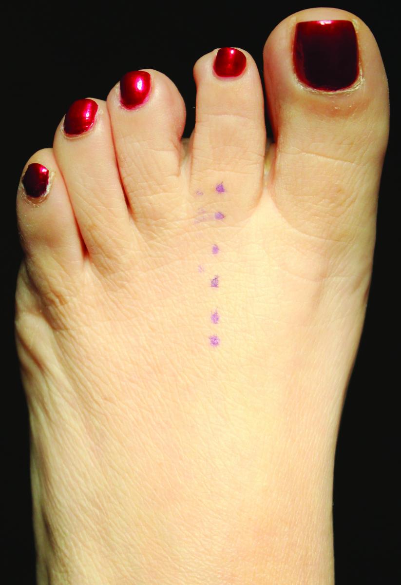
 Two posters that were recently presented at the American College of Foot and Ankle Surgeons annual meeting advocate different approaches, dorsal or plantar, to plantar plate repair. How do the two approaches stack up?
A poster by Erin Klein, DPM, and colleagues focuses on 53 patients who underwent a dorsal approach plantar plate repair via a Weil metatarsal osteotomy approach. After a two-year follow-up, the poster authors note that patients had less pain and edema, and more stability and strength at the metatarsophalangeal joint (MPJ). Patient reported outcome measures in this cohort of patients were significantly higher than at baseline.
Another poster by Mark Prissel, DPM, and colleagues analyzed the plantar approach to plantar plate repair in 76 patients. They note via validated outcome measures that pain, disability and activity limitation had significantly improved postoperatively, and 72.4 percent of patients noted their affected toes were touching the ground when standing.
Dr. Klein notes the dorsal approach allows surgeons to perform an osteotomy at the same time as the ligament repair through the same incision. Additionally, the collateral ligaments are easily visible and one can easily repair any visible pathology. Dr. Prissel has performed plantar plate repair to adjacent MPJs through a single incision and says this approach is amenable to all of the various deformities that surgeons will encounter with this repair.
Dr. Prissel adds that the plantar approach is very helpful in patients with subluxation/dislocation of the flexor tendons, transverse plane deformity requiring wedge resection of the plate for correction, and patients who do not require a shortening osteotomy.
Dr. Klein, an Associate at the Weil Foot, Ankle and Orthopedic Institute, says the organization of the fibers differs between the dorsal plantar plate (longitudinal organization) and the plantar portion of the plantar plate (transverse). She says a plantar approach does not allow one to see more subtle tears of the dorsal fibers or any pathology of the dorsal fibers unless there is a complete tear.
Dr. Prissel, a Fellow at the Orthopedic Foot and Ankle Center in Westerville, Ohio, cites several advantages to a plantar approach. As he notes, direct visualization of the plate is key to understanding the geometry of the tear/attenuation to ensure proper debridement of diseased tissue and correction of deformity. Additionally, Dr. Prissel notes that with more severe deformities, the flexor tendons may be dislocated either medial or lateral to the metatarsal head. In these situations, Dr. Prissel says the plantar approach provides more direct access to realigning the flexor complex, which helps prevent recurrence.
The disadvantages of the plantar approach may include concern for the plantar wound and concern over prolonged non-weightbearing. However, Dr. Prissel cites clinical data identifying plantar wound problems in only 4.2 percent of the patients in the study. He also notes that his protocol calls for protected weightbearing at one week postoperative. Since foot and ankle surgeons more commonly operate on the dorsal foot, Dr. Prissel says there may be a small learning curve with the dissection for the plantar approach. Overall, though, he says the plantar approach is “relatively simple and provides good exposure to the pathology.”
Dr. Klein adds that the plantar approach to plantar plate pathology will likely delay the initiation of rehabilitation/physical therapy, which she calls critical to the success of the procedure.
Dr. Klein has found the dorsal approach to be useful in almost all patients with plantar plate pathology. She does speculate that a dorsal approach may not be the best if the surgeon is making multiple longitudinal incisions on the foot but adds that one can modify the incision to accommodate this.
Could Subtalar Arthrodesis Lead To Joint Degeneration?
By Brian McCurdy, Managing Editor
Although subtalar arthrodesis is effective for subtalar joint disease, a recent study in the Journal of the American Podiatric Medical Association notes the procedure could induce joint degeneration and limited ankle joint motion.
The study focused on 37 patients who had subtalar arthrodesis and an average follow-up of 9.2 years. The authors noted improvements in both the Short-Form Health Survey and the American Orthopaedic Foot and Ankle Society ankle hindfoot score. However, patients did experience degenerative changes in the talonavicular, calcaneocuboid, metatarsocuboid and ankle joints.
Doug Richie Jr., DPM, FACFAS, points out that the most important issue in the study is the demonstration of osteoarthritis in the midfoot joints nine years after isolated subtalar joint fusion. He says this finding is common to other studies. Dr. Richie also points to the patients’ loss of ankle joint motion after the arthrodesis, which the authors attribute to a change in talus declination as a result of the fusion.
Dr. Richie emphasizes that the study focused on patients who received a subtalar fusion because of previous trauma and the vast majority had sustained calcaneal fractures, which then led to degenerative joint disease of the subtalar joint. Since the study was retrospective and authors made no comparisons to the preoperative X-rays, it is unknown if 100 percent of the post-op changes were due to the fusion procedure or the trauma that started the whole process, notes Dr. Richie, an Adjunct Associate Professor within the Department of Applied Biomechanics at the California School of Podiatric Medicine at Samuel Merritt University in Oakland, Calif.
As Dr. Richie notes, surgeons commonly perform subtalar arthrodesis for isolated degenerative joint disease or osteoarthritis of the subtalar joint, and less frequently for flatfoot deformity in adults.
Hindfoot height is almost always affected by calcaneal fracture and Dr. Richie suggests this is the case for the majority of the patients who underwent fusion in the study. He suggests reduced hindfoot height is not the result of subtalar joint fusion.
Dr. Richie has not found significant change in ankle joint motion in the sagittal plane in patients undergoing subtalar joint fusion and notes that most patients already had limited motion due to the arthritic process. Dr. Richie says the study authors did not measure pre-op range of motion. Questioning the study methodology, Dr. Richie says that although researchers compared post-op findings to the contralateral foot, they should have made the comparison to the preoperative radiographs that show the influence of trauma causing the need for surgery.
How Effective Is Laser Debridement For Chronic Wounds?
By Brian McCurdy, Managing Editor
Laser debridement has a positive healing effect on chronic lower extremity wounds, according to an abstract to be presented at the Symposium on Advanced Wound Care Spring/Wound Healing Society (SAWC Spring/WHS) meeting this month.
Researchers note the wavelength of the Er:YAG laser is at the peak of the absorption spectrum of water and can vaporize tissue with minimal adjacent thermal injury. The randomized study focused on 20 patients, 12 with venous ulcers and eight with diabetic foot ulcers, who received either laser or sharp debridement weekly. The laser debridement resulted in a significant increase in the percentage of the surface debridement in both venous and diabetic ulcers as well as a significant decrease in pain for debridement of venous ulcers but not for diabetic ulcers.
Alexander Reyzelman, DPM, feels laser debridement may have potential for painful ulcers and wounds. As he notes, with laser debridement, the pain level is significantly lower in patients and those who cannot tolerate sharp debridement can tolerate laser debridement. Dr. Reyzelman cautions that not all ulcers/wounds are appropriate for laser. He says deeper ulcers and those with necrotic tissue will still need sharp debridement.
Laser debridement is more precise than sharp debridement performed by hand, offers Dr. Reyzelman, an Associate Professor in the Department of Medicine at the California School of Podiatric Medicine at Samuel Merritt University. In addition, he says physicians doing laser debridement can control the depth of the laser and this is reproducible from one operator/physician to another. Dr. Reyzelman notes the precision of depth and uniformity lessens the chance of injuring vital tissue. With sharp debridement, Dr. Reyzelman notes there is more variation between the techniques of different physicians.
On the con side, Dr. Reyzelman notes laser debridement is expensive and cannot penetrate frank necrosis. He says laser debridement is usually for ulcers/wounds that have buildup of fibrinous film and bioburden, and wounds that are more superficial or shallow in nature.
The SAWC Spring/WHS will be held April 13-17 in Atlanta. For more info, visit www.sawcspring.com .


