Does An Abnormal Metatarsal Parabola Predict Plantar Plate Pathology?
 An abnormal metatarsal parabola is a risk factor for second MPJ plantar plate pathology, according to a study presented as a poster at the American Podiatric Medical Association (APMA) Annual Scientific Meeting.
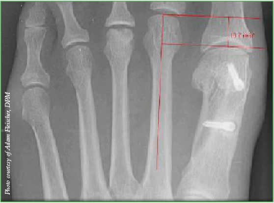
Researchers reviewed the records of 100 patients with a plantar plate injury and compared them to 200 patients in a control group. They found an abnormal metatarsal parabola, defined as a second metatarsal protrusion distance of more than 4 mm, was associated with almost 2.5 times the risk of developing plantar plate pathology. The authors note the study may help surgeons decide on the final resting position of corrective shortening of second metatarsal osteotomies.
Poster co-author Adam Fleischer, DPM, notes that more than a 4 mm difference in length between the first and second metatarsals on AP radiographs may suggest that a shortening metatarsal osteotomy of the second metatarsal, such as a Weil or other osteotomy, is indicated during second MPJ plantar plate surgery.
Dr. Fleischer and colleagues have found the most effective method for measuring the metatarsal parabola as it pertains to plantar plate pathology is Nilsonne’s method (see above X-ray). He acknowledges the limitation that this method may not work as well for patients with large intermetatarsal angles and the study’s findings may not be generalizable to all patients with various foot sizes. The average patient in the study was a 55-year-old female with a size 7 shoe, notes Dr. Fleischer, who is affiliated with the Weil Foot and Ankle Institute in Des Plaines, Ill. and is an Associate Professor of Radiology and Medicine at the Dr. William M. Scholl College of Podiatric Medicine.
Richard Bouché, DPM, points out that if the second metatarsal is too long and causing mechanical problems, then patients will have localized symptoms (pain, swelling and instability) to the second MPJ and clinical evidence of overload, which would include focal increased callus formation under the second metatarsal head. When treating the plantar plate, he notes one needs to address factors such as forefoot equinus and a tight Achilles. While Dr. Bouché is not opposed to a dorsal approach to plantar plate repair, he has experienced floating toe with this approach and prefers a plantar approach in his athletic population.
Dr. Bouché, a Fellow and Past President of the American Academy of Podiatric Sports Medicine, cites a variety of radiographic techniques and one clinical technique to validate metatarsal length abnormalities. He states the numerical angles or metric values obtained using different techniques are not comparable as measurement parameters are unique for each technique.
The best diagnostic sign of second MPJ plantar plate pathology, especially when positive, is a drawer test (the Lachman test), according to Dr. Fleischer. Dr. Bouché also cites the Lachman test but cautions against using it in isolation.
Dr. Fleischer notes a positive magnetic resonance image (MRI) is also highly specific for plantar plate tears and he rules it in when MRI is positive. On the flip side, he has found a negative ultrasound essentially rules out plantar plate injuries as the test is highly sensitive. Dr. Fleischer has also recently found that splaying of the second and third toes on AP radiographs can suggest a high-grade tear in the plantar plate.
To diagnose plantar plate pathology, Dr. Bouché also cites the efficacy of the pull-out test, when one slips a tongue depressor under the second metatarsal, and the Kelikian push-up test.
How Can Surgeons Ensure Post-Op Healing After TMAs?
By Brian McCurdy, Managing Editor
Surgeons should manage patient expectations before performing a transmetatarsal amputation (TMA), given that nearly half will not heal, according to a retrospective study presented as a poster at the APMA Annual Scientific Meeting.
Reviewing the cases of 27 patients with 29 transmetatarsal amputations, researchers found that 17 of the TMAs healed. There was a subsequent below-knee amputation in 10 patients and two patients died during follow-up. The study notes that surgeons had performed primary closure in 24 patients (14 with sutures and 10 with surgical staples) and left four TMAs open to heal by secondary intention.
Study co-author John Grady, DPM, notes that most factors that influence healing TMA procedures are the same factors affecting any procedures performed on patients with diabetes or infections: inadequate blood supply, elevated blood glucose levels, patient adherence, low serum albumin levels, poor nutritional status, obesity, immunosuppressive state, anemia and infection. He adds that surgeon-controlled issues negatively affecting healing include leaving inadequate flap coverage, placing too much tension on the incision, inadequate attention to the metatarsal parabola, and under-appreciation of the transverse plane level in attempting amputation.
As for adjunctive treatments that could speed healing, Dr. Grady cites concurrent vascular reconstruction or angioplasty, medical management (such as transfusions, antibiotic coverage, insulin and diet), skin grafting rather than closing tight incisions, and hyperbaric oxygen therapy.
Generally, if a TMA will not heal, it is time to consider a more proximal amputation, advises Dr. Grady, the Director of Podiatric Surgical Residency at the Jesse Brown Veterans Affairs Medical Center in Chicago. He says the amputation level is limited by factors such as inadequate blood flow or infectious disease process. In these cases, the other options are vascular reconstruction or getting to a level where blood flow is adequate to heal. One can then perform the amputation at the level with the best potential for healing and the least potential for destroying activity potential, according to Dr. Grady.
Can A New Hammertoe Fixation Device Improve On K-Wire Fixation?
By Brian McCurdy, Managing Editor
Fixation with a two-component implant with distal anatomic angulation was superior to K-wire fixation in patients with hammertoes, according to a recent study.
The prospective, randomized, multicenter study, presented at the recent American Orthopaedic Foot and Ankle Society (AOFAS) Meeting, focused on 58 patients. Half the patients had a standard arthrodesis with proximal interphalangeal joint resection and fixation with a K-wire while the other half had fixation via the Nextra Hammertoe Correction System (Nextremity Solutions). At six months, researchers noted that complete osseous union occurred in 83 percent of Nextra patients in comparison to 14 percent of those who had K-wire fixation.
Bradly Bussewitz, DPM, says advantages of the Nextra device include stability of the arthrodesis site in multiple planes as well as reduced pistoning risk. As he notes, adding stability should increase the chance of union and allow earlier weightbearing. Dr. Bussewitz says the internal nature of the device allows the patient to return earlier to regular shoe gear without the exposed K-wire and the lack of exposed K-wire should decrease the risk of infection.
With all the technological advances in the podiatric field, do K-wires still have a place for hammertoe or other lower extremity conditions? “The K-wire has withstood the test of time,” says Dr. Bussewitz, who feels the modality does have an active role in surgery.
Dr. Bussewitz, who practices at Steindler Orthopedic Clinic in Iowa City, Iowa, uses K-wires as well as metal and absorbable internal devices for hammertoe fixation. Implant costs are a factor in his decision making and anecdotally, he has experienced better fusion rates with K-wires than the 14 percent that this particular study described.
Although this AOFAS study only showed a 14 percent union rate with K-wires, Dr. Bussewitz says other studies have shown much higher rates of union with K-wires. He explains that factors like patient selection, surgical technique, perioperative protocol and differences in standards for assessing unions may be at play to explain the “vast difference” in union rates in the literature.
“A personal review of the literature leads me to believe successful outcomes are attainable with many different devices, including K-wires,” says Dr. Bussewitz.
Study: Naftifine Effective For Moccasin Tinea Pedis
By Brian McCurdy, Managing Editor
 A topical treatment scored high marks in a recent study of patients with moccasin tinea pedis.
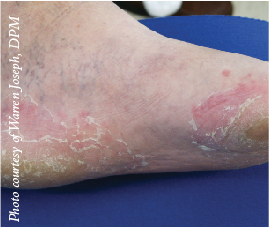
The data, presented as a poster at the APMA Annual Scientific Meeting, came from two double-blind, randomized multicenter studies. A total of 1,144 patients with either interdigital tinea pedis infection or interdigital moccasin tinea pedis received naftifine hydrochloride gel 2% (Naftin, Merz Pharmaceuticals) while 571 patients were in the vehicle group. Researchers found that pruritus, erythema and scaling improved significantly in the treatment group in comparison to the vehicle group. The poster notes that patients taking naftifine continued to experience improvements four weeks after treatment ended.
Patients tolerated the naftifine well, according to study coauthor Tracey Vlahovic, DPM. Noting that all prescription topical agents for tinea pedis only have approval for interdigital infection, she says the findings on moccasin tinea pedis could facilitate future studies.
“Even though this didn’t get specifically approved for moccasin tinea pedis, it opens the door for future research to define and study moccasin tinea pedis,” says Dr. Vlahovic, an Associate Professor and J. Stanley and Pearl Landau Fellow at the Temple University School of Podiatric Medicine.
What does this mean for future treatments for this condition? Dr. Vlahovic says the profession must first define to the FDA what moccasin tinea pedis is and then determine the duration and frequency of treatment. At this point, she notes physicians are unsure of the optimal treatment for this condition.


