ADVERTISEMENT
Current Insights On Neutral Casting And Biomechanical Exams For Orthotic Prescriptions
These expert panelists discuss the necessity of neutral casting for orthoses as well as what biomechanical exams to perform before prescribing an orthotic device.
Q:
Many people believe that the only requirement to make a good in-shoe orthotic is to take a neutral cast of the foot. Do you agree?
A:
 Taking a good cast is certainly an essential part of the process, according to Robert D. Phillips, DPM. If the cast is not correct, he asserts it is “extremely difficult if not impossible” to make an orthotic that will meet the patient’s goals and expectations.
By taking a cast of the foot in its “neutral position,” Dr. Phillips says one can fabricate an orthotic that starts pushing against the bottom of the foot when the shape of the foot matches that neutral shape you captured in the cast. However, he notes there are times when it is not desirable for the orthotic to push the foot toward a more neutral position.
Therefore, Dr. Phillips notes the importance of examining the patient to determine the pathology present, the abnormal forces and abnormal movements that are producing the pathology, and the source of the abnormal forces and movements. After determining these three factors, he says one can develop a plan that treats both the symptoms as well as the etiology of the symptoms. As Dr. Phillips points out, this treatment plan may involve a neutral orthotic, some other design of orthotic, some type of brace or physical therapy, or surgery.
Jarrod Shapiro, DPM, says if one defines “good in-shoe orthotic” as a device that alters the function of the foot and successfully improves patient symptoms, subtalar neutral casting is not enough. For example, he notes if the physician’s goal is to reduce stress on a tissue, then one must understand the rotational forces about the foot, which he says would allow the physician to write an appropriate prescription to modify the orthotic.
From this standpoint, Dr. Shapiro emphasizes an awareness that most people do not function in normal gait at the subtalar neutral position so placing the foot in neutral position may not be necessary or an appropriate primary goal. Additionally, he notes foot orthoses function to alter kinetic parameters rather than kinematic positions of bones.
Robert Eckles, DPM, notes that a comprehensive exam is essential in prescribing an effective orthosis. As he says, “You can’t know what you hope to capture if you do not” perform an exam. He cites variations to “neutral casting” that make sense in some scenarios and not in others. While rearfoot position is important and one should take care to approximate the neutral position, Dr. Eckles says proper positioning of the forefoot probably overshadows the importance of the rearfoot position.
“Failure to prepare the first metatarsal adequately, pronate the midtarsal joint and adequately dorsiflex the ankle are likely more important determinants of the function and acceptance of the device than simple rearfoot positioning,” asserts Dr. Eckles.
To attain a good orthotic, Scott Spencer, DPM, says not only is a good cast or impression of the patient’s foot essential, but one must also add features to influence and improve how the foot moves and interacts with the ground. He notes these features may include rearfoot posting, forefoot posting, a medial heel skive and a reverse Morton’s extension.
Q:
What type of evaluation should precede prescribing an orthotic for the shoe?
A:
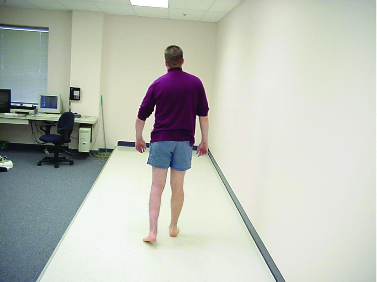
Dr. Eckles supports the idea of examining patient biomechanics, including stance and gait. He also advocates reviewing the patient’s age, medical history, height, weight, occupation stress, sport history and goals for treatment. As Dr. Eckles notes, these factors will determine how aggressive the prescription may be, whether the patient requires a surgical assist for correction, and clarify if the patient actually needs an ankle-foot orthotic (AFO) in lieu of a standard in-shoe device.
Dr. Phillips also advises performing a complete biomechanical examination, which includes range of motion as well as muscle testing and gait analysis. If the initial range of motion examination does not satisfy him in answering the questions as to why the patient is developing pathology, Dr. Phillips examines further factors, such as examination for abnormal subtalar joint axes or abnormal midtarsal joint axes. He notes that he cannot always come up with an immediate answer for every patient’s symptoms and sometimes must be innovative in his solutions.
Dr. Shapiro’s orthometric examination, rather than measuring specific joint motions in degrees, includes a sitting and standing examination of the entire limb. He will also measure rearfoot position, joint motion, location of the subtalar joint axis in the transverse plane, the forefoot to rearfoot relationship, and specific provocative tests of the affected tissue depending on the patient complaint. Dr. Shapiro will also perform a visual gait analysis.
“I am looking to appreciate the significant biomechanical interactions affecting my patient’s diagnosis,” says Dr. Shapiro. “For example, hallux valgus requires a full examination to determine the mechanical contributions to the bunion rather than just focusing on the bump.”
Dr. Spencer advises clinicians to assess the patient’s current shoes and find out the styles of shoe the patient is likely to wear. If the patient’s choice of shoe is not conducive to working with a foot orthotic device, he says one should relay the importance of changing shoe gear to the patient before prescribing an in-shoe foot orthotic device. Dr. Shapiro also emphasizes the importance of observing the wear pattern on the outsole of the shoe. Gait analysis with and without patients wearing shoes can provide information on what an orthotic for that patient will need to do to improve gait function, according to Dr. Spencer.
Dr. Phillips says one must find out if the patient really does need a custom-made orthotic or whether there is a prefabricated orthotic out there that can still meet the patient’s goals. Therefore, he notes a complete examination of the patient, entailing a good history and physical exam, “is our moral obligation.”
“I don’t believe that we should take any less care in evaluating the patient for orthotics than we do in evaluating patients for surgery. That’s what the patient is paying for,” says Dr. Phillips. “If patients want arch supports, they can go to their local drugstore, step on a machine that has some predetermined paradigms in it and get an arch support that may be very effective in doing what the patient is looking for.”
Q:
What type of biomechanical exam would you advocate to decide what type of flatfoot or cavus foot surgery to do?
A:
There is no “one size fits all” answer for Dr. Spencer. He says stance measurements such as neutral calcaneal stance position and resting calcaneal stance position, even though they are not reflective of the actual effect on the foot by ground reactive forces on the foot, can give an idea of the status of the weightbearing foot. To understand the implications of a procedure and possible effect on the patient’s post-op gait, Dr. Spencer also cites using several measurements, including ranges of motion of the first metatarsophalangeal joint, midtarsal joint, subtalar joint and ankle joint.
Dr. Eckles will perform a full biomechanical examination, including gait analysis. For flatfoot or cavus procedures, characteristics such as planal dominance, superstructure alignment and velocity of motion are all too important and easy to overlook. Since flatfoot and cavus foot surgery will likely result in deliberate restriction of motion at one or more joints, Dr. Eckles says a biomechanical evaluation is important not only to identify the mechanical etiology (site and contributors) of the deformity, but to plan for the position, motion and compensations that the post-op foot will demonstrate.
Dr. Shapiro does not change his biomechanical exam for flatfoot or cavus patients, but notes performing additional exams for those conditions. For example, he says surgery for a rigid flatfoot is much different from that of a flexible flatfoot so he would perform tests such as the supination lag test to determine the integrity of the spring ligament. Dr. Shapiro would also evaluate the position of the forefoot when placing the rearfoot into a corrected position as well as midfoot mobility. As he says, these exams help in determining what forefoot surgical procedure is necessary.
If the patient has a relaxed calcaneal stance in which the heel is vertical but with an extremely flat arch, Dr. Phillips says flatfoot surgery may leave the rearfoot in a varus state, creating more problems for the patient than before surgery. Therefore, he notes the importance of stance measurements that would calculate not only the current function of the rearfoot but the function of the rearfoot postoperatively as well.
Dr. Phillips also advises caution when doing surgery for compensation in cases that prohibit control of the causes of the compensation. For example, if one performs surgery on a patient with a very flat foot with high internal tibial torsion, he says surgery could theoretically cause the patient to walk pigeon-toed after surgery. Dr. Phillips notes a short iliopsoas muscle could be problematic if the patient has flatfoot surgery, saying the cause of the flatfoot is still very much there.
Dr. Phillips believes the biomechanical examination is more important before doing flatfoot surgery or cavus foot surgery than it is even before prescribing orthotics, citing the importance of examining abnormal subtalar and midtarsal joint axes. He also notes it is crucial to examine for abnormal forefoot to rearfoot relationships as well as calculating the post-op forefoot to ground relationships. Dr. Phillips notes evaluating the length of the Achilles post-op can ensure the patient will not have increased pronation forces after surgery.
For the cavus foot, Dr. Shapiro emphasizes understanding if there is a neurological component and what muscle strength remains in the specific muscle tendon units. For example, he notes a patient with Charcot-Marie-Tooth disease will have a posterior tibial tendon overpowering a weak peroneus brevis, which will become integral in determining which tendon transfers or augmentations are necessary. He adds that a subtalar fusion is unlikely to be successful if the surgeon does not consider the contribution of tibial varum to the position of the foot.
Dr. Eckles is the Dean of Clinical Studies and an Associate Professor in the Department of Orthopedics and Pediatrics at the New York College of Podiatric Medicine.
Dr. Shapiro is an Associate Professor with the College of Podiatric Medicine at the Western University of Health Sciences in Pomona, Calif. He is the Director of the Chino Valley Medical Center PMSR/RRA Podiatric Residency in Pomona, Calif.
Dr. Spencer is an Associate Professor Surgery/Biomechanics at the Kent State University College of Podiatric Medicine. He is a Fellow of the American College of Foot and Ankle Orthopedics and Medicine.
Dr. Phillips is affiliated with the Orlando Veterans Affairs Medical Center in Orlando, Fla. He is a Diplomate of the American Board of Foot and Ankle Surgery, and the American Board of Podiatric Medicine. Dr. Phillips is a Professor of Podiatric Medicine with the College of Medicine at the University of Central Florida. He is also a member of the American Society of Biomechanics.
For further reading, see “Addressing Recent Controversies And Developments In Orthotic Therapy” in the August 2017 issue of Podiatry Today, “Key Pearls On Orthotic Casting And Fabrication” in the December 2016 issue or “Ensuring Patients Get A Perfect Orthotic Fit” in the October 2015 issue.


