Could Micro-Autografts Have Potential In Healing Diabetic Foot Ulcers?
The goal of wound management is to attain cost effective wound closure to reestablish function.1 This is especially true with lower extremity wounds that are debilitating and often lead to a temporary or permanent disability.
For those lower extremity wounds we cannot close in a primary manner, other options are spontaneous closure by secondary intention or tertiary closure with a skin graft, pedicle or free flap. Spontaneous closure by a combination of wound contraction and epithelialization often results in an unstable wound that is prone to recurrent breakdown. Researchers have noted that autologous wound closure is the gold standard for wound closure.2
Studies have demonstrated that the use of split-thickness skin grafts (STSGs) to close wounds such as diabetic foot ulcers and venous ulcers can decrease the time to healing, minimize morbidity and decrease the cost of care.2,3-6 Mahmoud and colleagues reported that diabetic foot ulcers treated with STSG healed in 28 days versus 122 days for conservative wound care.3 If one is using STSG to close lower extremity wounds, it is important that such grafts include both the dermis and the epidermis.
There are several distinct advantages of transplanting dermis into the wound. The amount of wound contraction that occurs in a grafted wound is directly proportional to the amount of dermis in the graft.7,8 The fibroblasts in the mesenchymal mesodermal tissue of the dermis produce high levels of growth factors that facilitate the proliferative phase of healing. Grafts with dermis contain elevated levels of tumor necrosis factor-alpha (TNF-a), platelet-derived growth factor (PDGF) and fibroblast growth factor (FGF).9,10 All of these factors favor re-epithelialization, neoangiogenesis and extracellular matrix deposition. This advantage is not present in epidermal grafts that only contain keratinocytes.11
Finally, grafts containing dermis provide improved tensile strength to the repaired wound. This is especially important in wounds on the sole of the foot. Grafts containing only epidermis are not stable and provide very limited strength. The history of cultured epithelial autografts demonstrates a fragile nature and susceptibility to blistering and shearing.11
A Closer Look At An Innovation In STSG Harvesting
Recently, a device has emerged that allows one to harvest a very small STSG of medium thickness in an outpatient procedure with local anesthesia. The Xpansion micro-autografting kit (SteadMed Medical) also allows expansion of a postage stamp-sized graft up to 100 times the size of the donor site.12 The kit includes a disposable hand-powered, precalibrated dermatome that produces uniform 0.012- to 0.016-inch thick STSGs. It also contains a mincer that cuts the graft into fragments of 0.8 x 0.8 mm in size. These fragments contain both dermis and epidermis, rete ridges or pegs, and one can place the fragments onto the wound without concern for orientation.12,13
We have used this outpatient technique for grafting in our clinic for the past year with great success. We will present three case examples of Xpansion micro-autografting. In each of the cases, we attempted standard wound care to encourage spontaneous healing. When permanent stable healing did not occur, we proceeded to utilize Xpansion micro-autografting with minced skin grafts as an outpatient procedure. In each case, graft take occurred and the wound remained healed.
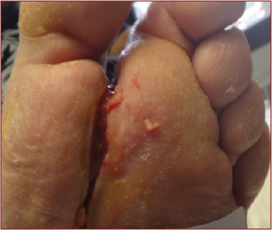
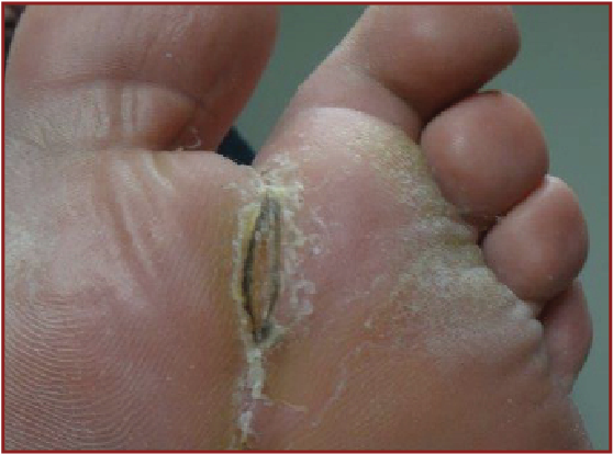
 Case 1. A 40-year-old Caucasian male presented with a chronic diabetic ulcer in the first metatarsal space. After sharp debridement and the use of hypochlorous acid soaks and hyperbaric oxygen (HBOT), the ulcer slowly closed by secondary intention over the next six weeks. However, seven days after healing, the wound closure broke down and essentially left the original wound. After a week of wound bed preparation, we performed micro-autografting as an outpatient procedure with local anesthesia. The posterior calf was the donor site. The wound was completely healed 14 days later and has remained healed for the past six months.
Case 2. A 50-year-old Hispanic male presented with a chronic infected diabetic ulcer on the medial lower leg. In addition to getting oral antibiotic treatment, the patient received topical Silvadene Cream and Bactroban ointment to eradicate the infection. We performed Xpansion micro-autografting as an outpatient procedure with local anesthesia. The posterior calf was the donor site. Two weeks later, the spreading skin fragments were visible in the wound. After another three weeks, the wound was totally healed in both dermis and epidermis.
 Case 3. A 65-year-old African-American male presented with a history of deep vein thrombosis, chronic edema and cellulitis in a leg with a venous stasis ulcer. He also had an abscess that required incision and drainage in the operating room. Conservative treatment with compression therapy, weekly debridement and Regranex (Smith and Nephew) therapy over the next 14 weeks did not result in total wound closure. Subsequent micro-autografting in the outpatient clinic lead to complete closure of the wound. The posterior calf was the donor site. At two weeks, all grafts had taken and were visibly coalescing. After an additional week, we removed the keratin and a healed wound was evident.
Additional Insights And Study Findings On Micro-Autografting
The three cases demonstrate the ease of achieving permanent wound closure (in the dermis and epidermis) through an outpatient procedure with local anesthesia. The key to success with this technique is optimal wound bed preparation. One must remove all necrotic tissue and debris from the wound. The wound bioburden must be minimal and one must remove deleterious cytokines such as excessive matrix metalloproteinases from the wound.14 After achieving optimal wound bed preparation, clinicians can attain autologous skin closure with epidermis and dermis without hospital admission, thereby conserving costs, time and resources.12
In a large case series, Boggio and colleagues reported that micrografts induced faster reepithelialization of chronic leg ulcers that had failed to heal despite good conservative local therapy.15 They also stated that they could repair very large ulcers with small fragments of skin requiring small donor sites. Boggio and coworkers noted a 90 percent success rate with the micrografting technique.
In a controlled experimental study, researchers compared micrografted wounds to control non-grafted wounds.16 The micrografted wounds were 80 percent healed by 10 days and 100 percent healed by 14 days in comparison to 40 percent of non-grafted control wounds healed at 10 days and 60 percent healed by 14 days.
Final Notes
I have found that micro-autografting using the Xpansion kit is an excellent method to obtain permanent wound closure of lower extremity wounds. The speed of wound closure after micro-autografting is very gratifying for patients, who have wounds for a prolonged period of time, as well as clinicians, who find much faster healing rates using this technique.
Dr. Caputo is Director of the Wound Care Center at Clara Maass Medical Center in Belleville, N.J.
Dr. Fahoury is the Chief of Podiatry at Monmouth Medical Center in Long Branch, N.J.
References
- Tobin GR. Closure of contaminated wounds: Biologic and technical considerations. Surg Clin N Amer. 1984; 64(4):639-52.
- Anderson JJ, Wallin KJ, Spencer L. Split thickness skin grafts for non-healing foot and leg ulcers in patients with diabetes: A retrospective review. Diabetic Foot Ankle. 2012; Epub Feb 20.
- Mahmoud SM, Mohamed AA, Mahdi SE, Ahmed ME. Split-thickness skin graft in the management of diabetic foot ulcers. J Wound Care. 2008; 17(7):303-306.
- Rosenblum B. Berglund A. Split thickness skin grafts used for diabetic wound healing. Podiatry Management. 2012; 31(5):141-146.
- Ramaanujam CL, Stapleton JJ, Kilpadi KL, Rodriguez RH, et al. Split-thickness skin grafts for closure of diabetic foot and ankle wounds: A retrospective review of 83 patients. Foot Ankle Spec. 2010; 3(5):231-240.
- Wood MK, Davies DM. Use of split-skin grafting in the treatment of chronic leg ulcers. Ann R Coll Surg Engl. 1995; 77(3):222-223.
- Wood BC. Skin grafts and biologic skin substitutes. Emedicine. Available at https://emedicine.medscape.com/article/1295109-overview .
- Harrison CA, MacNeil S. The mechanism of skin graft contraction: An update on current research and potential future therapies. Burns. 2008; 34(2):153-163.
- Pertrusi G, Tiberio R, Graziola F, Boggio P, et al. Selective release of cytokines, chemokines, and growth factors by minced skin in vitro supports the effectiveness of autologous minced micrografts technique for chronic ulcer repair. Wound Rep Regen. 2012; 20(2):178-184.
- Sharma K, Bullock A, Ralston D, MacNeil S. Development of a one-step approach for the reconstruction of full thickness skin defects using minced split thickness skin grafts and biodegradable synthetic scaffolds as a dermal substitute. Burns. 2014; 40(5):957-965.
- Desai MH, Mlakar JM, McCauley RL, Abdullah KM, et al. Lack of long-term durability of cultured keratinocyte burn wound coverage: A case report. J Burn Care Rehab. 1991; 12(6):540-545.
- Smith DJ. Achieving efficient wound closure with autologous skin. Today’s Wound Clinic. 2014; 8(1):23-24.
- Svensjo T, Pomahac B, Yao F, Slama J, et al. Autologous skin transplantation: Comparison of minced skin to other techniques. J Surg Res. 2002; 103(1):19-29.
- Robson MC. Advancing the science of wound bed preparation for chronic wounds. Ostomy Wound Manage. 2012; 58(11):10-12.
- Boggio P, Tiberio R, Gattoni M, Colombo E, Leigheb G. Is there an easier way to autograft skin in chronic leg ulcers? “Minced micrografts”, a new technique. J Eur Dermatol Venereol. 2008; 22(10):1168-1172.
- Hackl F, Bergmann J, Granter S, Koyama T, et al. Epidermal regeneration by micrograft transplantation with immediate 100-fold expansion. Plast Reconstr Surg. 2012; 129(3):443e-452e.
For further reading, see “Essential Insights On Surgical Management Of Diabetic Foot Ulcers” in the December 2014 issue of Podiatry Today or “Assessing The Evolution Of Advanced Products For Diabetic Foot Ulcers” in the November 2014 issue.
For an enhanced reading experience, check out Podiatry Today on your iPad or Android tablet.


