ADVERTISEMENT
A Closer Look At Simple Surgical Solutions For Diabetic Foot Ulcers
 Ulcers of the toes and forefoot in patients with diabetes are sometimes difficult to heal or keep healed. Even with good local wound care, appropriate offloading during wound healing, extra-depth shoes and/or custom offloading orthotics, and regular follow-up care, patients often reulcerate, particularly in the presence of neuropathy and structural foot deformity.
It is of course important to rule out and address other reasons for non-healing or recurrent ulceration such as peripheral arterial disease, poor control of diabetes, malnutrition, smoking or infection.1 Ideally, healing of an ulcer is preferable prior to any surgical intervention but sometimes surgery is required to offload the area adequately to achieve healing.
Obviously, some patients require surgical intervention regardless of their “fitness” for surgery when significant infection is present. However, when it comes to elective procedures, we must choose our patients carefully. Those patients who have not been adherent with management of their ulceration may not be good candidates for surgery. A full workup of the patient and involvement of other medical specialties to maximize the management of all comorbidities are necessary prior to surgical intervention.
The surgeon should also be aware of social factors that might prohibit success such as lack of help at home, lack of transportation to get to postoperative visits, the need for assistive devices to aid mobility at home and outside the home, etc. One can use a local anesthetic for many forefoot procedures to reduce the perioperative risk to patients, especially if they have significant cardiovascular comorbidities.
When There Is A Recurrent Hallux Interphalangeal Joint Ulceration
A recurrent hallux interphalangeal joint ulcer can heal with appropriate offloading and local wound care, but it is often very difficult to keep it healed despite the use of offloading orthotics and shoe gear.
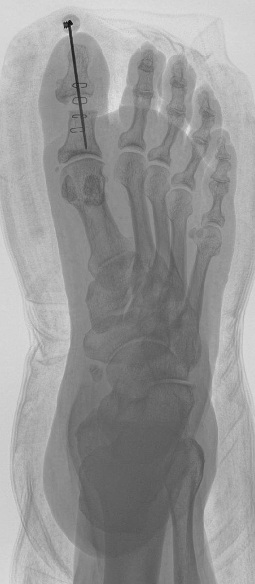
An effective treatment is a hallux interphalangeal joint arthroplasty. Perform a linear incision over the hallux interphalangeal joint. Deepen the incision to the level of the extensor tendon and transect the tendon at the joint line along with the collateral ligaments. Expose the head of the proximal phalanx and resect it with a sagittal saw. Drive a 0.062 Kirschner wire from the base of the distal phalanx out the end of the hallux and then retrograde the K-wire back through the proximal phalanx, holding the hallux in a straightened position.
Leave the K-wire in for three weeks and remove it in the clinic.2 Patients can bear partial weight on the right heel in a surgical shoe with either a cane or walker for stability. If they are unable to keep partial weight on the heel, they may just take shorter steps, placing the surgical foot forward first and bringing the unoperated foot up to meet it, rather than surpassing it, in order to avoid rolling over the toes on the operated side.
This procedure can also be useful for a hallux interphalangeal joint contracture causing distal or dorsal pressure in shoes for a patient who is not a good candidate for a fusion.
I have also had a few patients who had partial hallux amputations at the interphalangeal joint without removal of the proximal phalangeal head. In two of these patients, ulcerations were developing plantar to the head of the proximal phalanx and were difficult to heal and keep healed. Resection of the head of the proximal phalanx through a dorsal linear incision was effective in getting and keeping the ulcers healed. In general, one should always remove the head of the proximal phalanx in a partial hallux amputation when removing the entire distal phalanx to help avoid this complication.
How To Resolve A Recurrent Distal Lesser Toe Ulcer In The Presence Of A Flexible Hammertoe
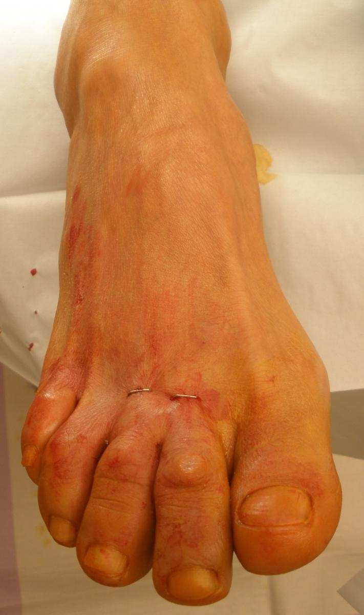
For a recurrent distal ulcer in the lesser toe in a patient with flexible hammertoe, perform a percutaneous extensor and flexor tenotomy to straighten the toe and eliminate pressure on the distal end of the digit.
 One can perform this with the patient having a local anesthetic. Generally, I perform both a flexor and extensor tenotomy to avoid excessive dorsiflexion at the metatarsophalangeal joint (MPJ) if the flexor tenotomy occurs in isolation.
Utilize a 64 beaver blade for this procedure. Make the initial stab incision dorsal to the MPJ in a longitudinal fashion. Then deepen the blade to the level of the joint and rotate the blade medially and laterally, cutting the extensor tendon and joint capsule at that level. Plantarflex the toe at the MPJ to spread the tendon ends apart.
Then direct attention to the plantar aspect of the proximal interphalangeal joint of the toe (located at the toe sulcus). Make a linear incision with the 64 blade, deepening it to the level of the joint. Rotate the blade medially and laterally, cutting the flexor tendon and joint capsule at this level. Dorsiflex the toe to spread the cut ends of the flexor tendon apart. If adequate correction has not yet occurred, make an additional stab incision plantar to the distal interphalangeal joint of the digit and perform the identical procedure there. If the patient only has a mallet toe, the surgeon may only need to perform a single procedure plantar to the distal interphalangeal joint.
Following adequate release, close the skin with a single skin staple at each stab incision.3 Dress the surgical site with betadine-soaked gauze for splintage around the toes generally for two to three weeks. During this time, the patient remains partially weightbearing on the heel in a surgical shoe. Remove the skin staples and the patient returns to normal shoe gear. Inform patients preoperatively that they will not be able to grasp with the operated toes after surgery.4
Key Insights On Addressing A Plantarflexed Lesser Metatarsal Head With Recurrent Ulceration
A percutaneous elevating osteotomy without fixation can be an effective treatment for a plantarflexed lesser metatarsal head with a recurring ulcer.
To identify proper placement of the surgical incision, place a thumb under the plantar aspect of the metatarsal head and the index finger of the same hand at the dorsal aspect of the metatarsal in line with the thumb. This allows you to identify the location of the surgical neck or the level of the metaphyseal-diaphyseal junction at which one will perform the osteotomy. The skin incision will be located just proximal to the index finger at the surgical neck of the metatarsal in a transverse fashion, extending about 1 cm in length.
 Use a hemostat to dissect down to the level of the metatarsal. Then open the jaws and place the hemostat on either side of the metatarsal from dorsal to plantar. This will move the extensor tendon to one side and enable one to dissect the soft tissues away from the dorsal, medial and lateral aspects of the bone. It is not necessary to make a periosteal incision unless you need to resect a portion of bone to shorten the metatarsal. One can replace the desired hemostat with Hohmann retractors prior to making the osteotomy. Under radiological guidance, use a sagittal saw to make an osteotomy at the surgical neck of the metatarsal from slightly dorsal proximal to plantar distal. This will allow dorsal and proximal translation of the capital fragment. An osteotome can verify completion of the osteotomy. You may grasp the capital fragment dorsally and plantarly as you did to identify the location of the skin incision, and move the capital fragment dorsally and plantarly to ensure complete release.
No fixation is necessary. Stabilize the metatarsal head by the deep transverse intermetatarsal ligament, joint capsule and ligaments. No deep closure is necessary. Just close the skin incision. I recommend using a dressing with extra padding under the osteotomized metatarsal head to maintain dorsal translation of the metatarsal head until skin healing. The patient bears partial weight in a surgical shoe during bone healing.5,6
In Conclusion
Although patients with diabetic ulcerations can often have multiple comorbidities (sometimes with poor management), one can often perform the aforementioned procedures, even with the utilization of a local anesthetic, to help eliminate the deforming forces that are preventing healing or causing repeated recurrence of the ulceration. This can allow for improved quality of life in this patient population and help avoid amputation. Still, one must carefully select appropriate patients for these procedures. Any patient who has had a previous ulceration, even after deformity correction, will require long-term follow-up with appropriate shoe gear, orthotics and possibly bracing to help maintain the health of their lower extremities.
Dr. Schweinberger is affiliated with the Veterans Affairs Medical Center in Cheyenne, WY. She is a Fellow of the American College of Foot and Ankle Surgeons.
References
- Schweinberger MH, Roukis TS. Wound complications. Clin Podiatr Med Surg. 2009;26(1):1-10.
- Lew E, Nicolosi, N, McKee P. Evaluation of hallux interphalangeal joint arthroplasty compared with nonoperative treatment of recalcitrant hallux ulceration. J Foot Ankle Surg. 2015;54(4):541-548.
- Landsman A, Cook E, Cook J. Tenotomy and tendon transfer about the forefoot, midfoot and hindfoot. Clin Podiatr Med Surg. 2008; 25(4):547-69.
- Debarge R, Philippot R, Viola J, Besse JL. Clinical outcome after percutaneous flexor tenotomy in forefoot surgery. Int Orthop. 2009; 33(5):1279-82.
- Schade VL, Roukis TS. Minimum-incision metatarsal osteotomies. Clin Podiatr Med Surg. 2008; 25(4):587-607.
- Lui TH. Percutaneous dorsal closing wedge osteotomy of the metatarsal neck in management of metatarsalgia. Foot (Edinb). 2014;24(4):180-5.
Editor’s note: For further reading, see “The Multidisciplinary Team Approach To The Diabetic Foot” in the June 2016 issue of Podiatry Today, “Essential Insights On Surgical Management Of Diabetic Foot Ulcers” in the December 2014 issue or “Emphasizing The Fundamentals And Patient Education In Diabetic Foot Care” in the September 2014 issue.
To sign up for Podiatry Today’s weekly e-newsletter, visit www.podiatrytoday.com/e-news.


