ADVERTISEMENT
A Closer Look At Prophylactic Surgery In Patients At High Risk Of Ulceration
 Historically, prophylactic surgical offloading in patients with diabetes has come with little recommendation. However, more recent studies have acknowledged its importance in the management of recurrent or longstanding ulcerations.1-3 In fact, surgical procedures often present as viable or even the most beneficial course of therapy in select patients with diabetic foot ulcerations (DFU).
Many non-surgical offloading modalities exist to promote the healing of a DFU and prevent recurrence. However, no treatment or protective measure will ever be 100 percent effective. For example, prescription therapeutic shoe gear, which may arguably be the mainstay in the regimen for attempted prevention of primary and recurrent DFUs, is limited by patient adherence.1,4 Researchers have demonstrated that approximately one in three patients are unable to maintain adequate adherence to recommended use guidelines.4 Even with the best efforts of clinicians practicing non-surgical wound prevention therapies, researchers have reported annual recurrence rates of DFUs as high as 34 percent, 61 percent and 70 percent at one, three and five years respectively, and some recurrence rates were as high as 20 to 58 percent within one year.5,6
As the search for increasingly effective non-surgical prevention modalities continues, physicians should remain diligent in observing and addressing ulcer promoting deformities and forces that one may permanently “offload” with surgical intervention.
Keys To The Clinical And Perioperative Evaluation
Scrutiny is paramount when it comes to appropriate patient and procedure selection for surgical intervention in an effort to reduce or prevent ulcerations. Patients with complications of diabetes, including peripheral neuropathy, are at a tenfold increased risk of postoperative complications.7 Accordingly, one must thoroughly review and evaluate non-surgical options prior to discussing surgical intervention.
Suitable patients for prophylactic surgery are those with otherwise controlled comorbidities, have demonstrated adherence to therapeutic recommendations and have a history of appropriately maintaining a follow-up schedule. One can evaluate long-term glucose control with the hemoglobin A1c (HbA1c) test. A HbA1c >7% is directly related to poor post-surgical outcomes including soft tissue infection, bone healing complications and prolonged healing in comparison to patients with a hemoglobin A1c <7%.8-10 At a minimum, clinicians should emphasize short-term glucose control to patients as elevation of glucose levels leads to protracted healing and increased incidence of dehiscence.11,12 Additionally, one should perform basic vascular exams, including an ankle brachial index (ABI), which is recommended for all patients with diabetes greater than 50 years of age.13
 Guidelines from the American Diabetes Association (ADA) suggest follow-up specialist visits every three to six months for patients with a foot deformity and loss of protective sensation, and every two months for those with a history of ulceration or amputation. These office visits are of ideal frequency to routinely evaluate the need for prophylactic surgery.14 Clinical findings that may indicate surgical consideration include recurrent hyperkeratotic lesions, erythema, increased temperature in comparison to contralateral limbs, intradermal hemorrhage and soft tissue erosions.15 Additional exams to aid in surgical procedure selection include weightbearing radiographs, a brief gait exam and biomechanical evaluation including the first MPJ, lesser digit range of motion (ROM) and ankle ROM. Evaluation for appropriate fit, function and physical state of shoe gear is also standard.
What To Consider In The Pathomechanical Evaluation
Often, the clinician can readily observe the pathophysiology of recurrent ulceration while performing a basic biomechanical and musculoskeletal evaluation. A patient with recurrent dorsal proximal interphalangeal joint ulcerations and rigid hammertoe deformity may benefit from a hammertoe correction procedure. In other cases, one should give more consideration to more proximal pathology, such as first ray insufficiency, bunion deformity, tendon contracture or equinus deformity, which may be contributing to ulceration. As many of these pathologies are secondary to the long-term course of diabetes, an appropriate surgical procedure is one that considers or corrects all contributing forces.7,16
In all cases of DFU, clinicians should evaluate the ankle for an equinus deformity as less than 10 degrees of dorsiflexion at the ankle is often a contributing factor to forefoot ulcerations.17 Ankle equinus is often a contributing factor with recurrent DFUs. When patients demonstrate an equinus deformity of the gastrocsoleus complex, one can easily perform a tendo-Achilles lengthening, which will facilitate pressure reduction and offloading of the forefoot during gait in neuropathic patients.17
Addressing Recurrent Ulcerations Adjacent To The First Metatarsal Head
Considerations for recurrent ulceration adjacent to the first metatarsal head include prominences due to metatarsal positioning in the transverse and sagittal planes, sesamoid position or prominence, and arthritic changes to the first MPJ.
 Recurrent ulceration to the plantar metatarsal head should be an indication for scrutiny of the metatarsal declination angle or sesamoid prominence due to fat pad atrophy or ankylosis of the flexor apparatus. When lesions present to the lateral base or purely to the lateral aspect of the hallux, a thorough workup for hallux valgus or first MPJ pathology is in order.
One may remove bony prominences, including sesamoids, with a simple, carefully planned osteotomy or resection. In cases of recurrent ulceration to the plantar first metatarsal head after sesamoidectomy or those cases in which osteomyelitis is present in the metatarsal head, the clinician may perform a total first joint resection. This procedure, as described by Frykberg and colleagues, should include the removal of the metatarsal head, the base of the proximal phalanx and both sesamoids.18 Retention of the sesamoids, as related by the authors, can create a fibrous bridge with the metatarsal stump and become a source for further ulcer recurrence.
While assessing the forefoot to rearfoot relationship and evaluating first ray position, one may note a plantarflexed first metatarsal head secondary to pathologic tension of the peroneus longus tendon. Recommended treatment for this soft tissue driven cavus foot deformity is tenodesis of the peroneus longus to the peroneus brevis.19
The surgeon may address a rigid deformity with the use of an osteotomy designed to elevate the metatarsal head or by performing a partial first metatarsal head excision. If a procedure to correct hallux valgus deformity or the metatarsal declination angle is desired, one must bear in mind that patients with diabetic neuropathy also demonstrate higher levels of non-union or malunion. Although surgeons often perform these procedures with great success, this topic remains controversial.
Patients with fat pad atrophy or decreased soft tissue secondary to recurrent ulceration may benefit from soft tissue augmentation. Historically, silicone injections were available. Currently, autologous fat pad grafting is gaining popularity. First described in 1994 by Chairman, the “autolipotransplantation” fat grafts to the plantar foot provided good results in 50 patients.20 More recent evaluation indicates that fat grafting provides improved plantar load distribution as well as local soft tissue stability.21 Luu and colleagues have provided a detailed outline of this process and demonstrate recurrence prevention in a single patient.22
Pertinent Insights On Procedure Selection For DFUs At The Hallux
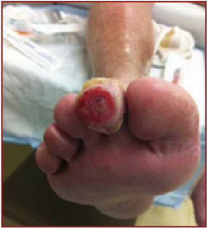
The hallux is a common anatomic area for recurrent DFUs with a more proximal reamputation reportedly occurring in as frequently as half of those patients who have any partial first ray resection.23-25 One should carefully evaluate for clinical evidence of pre-ulcerative lesions or etiologies of recurrent ulcerations to all aspects of the hallux when treating these patients.
Medial hallux wounds are frequently secondary to biomechanical abnormalities that transfer excessive force to the medial aspect of the hallux phalanges. This results in soft tissue changes including hyperkeratosis or intradermal petechiae, which can often progress to ulceration. For each patient with a medial hallux soft tissue wound, clinicians should evaluate for pronatory motion or position in gait, medial column insufficiency, hallux valgus and hallux rigidus deformities. When accommodative and supportive measures fail, one should consider therapeutic surgical intervention.
Plantar hallux wounds may be secondary to bony exostoses or os interphalangeus sesamoids. Often, first MPJ pathology, such as hallux limitus, at least partially contributes to these wounds, leading to increased pressure distally. A carefully selected exostectomy or sesamoidectomy procedure is indicated for isolated pathologies or in conjunction with other procedures when there are combined pathological states.
The Keller procedure, or the first MPJ arthroplasty by surgical resection of the base of the proximal phalanx, has been a surgical mainstay for nearly half a century in patients with first MPJ rigidity or hallux valgus deformity. The addition of indications for patients with diabetes, neuropathy and first MPJ rigidity causing recurrent ulceration has augmented the therapeutic spectrum of the Keller procedure.26 Armstrong and colleagues have demonstrated a significantly increased rate of wound healing and a decreased rate of ulcer recurrence to the hallux in comparison to non-surgical treatment of patients with diabetic neuropathy.27 Common post-op complaints after the Keller procedure include decrease in propulsive force and decrease in hallux purchase with occasional floating hallux.
 Interphalangeal joint arthritis, extensus and malleus also cause a pathologic increase of plantar pressures to a relatively small area of tissue and focal areas of ulceration to the plantar or distal hallux. The surgeon should address any flexible soft tissue contractures with any of the well described tenotomy and skin plasty techniques. Patients with significant arthritis or rigid deformity require an osseous procedure such as arthroplasty or arthrodesis. Arthrodesis is historically indicated in patients with a more active lifestyle as they require increased stability of the hallux during the toe-off phase of gait.
When Procedures Involve The Central Rays
Motor neuropathy is a common finding in patients with diabetes but this is most commonly limited to the foot and more specifically to the intrinsic musculature of the foot. Decreased muscular function leads to atrophy, imbalance and deformity.28-30 One of the most observed deformities in the diabetic foot is digital contraction leading to hammered, clawed or mallet toe. Beginning as a soft tissue imbalance, hammertoe deformities progress to rigid positional deformities that are a frequent source of ulceration and soft tissue breakdown. Hammertoe deformities promote ulceration of the distal digit due to increased plantar pressures in gait as well as the dorsal digit due to friction from contact with shoe gear.
One can easily address flexible hammertoe deformities with soft tissue correction including flexor tenotomy or tendon lengthening and capsular procedures. Various authors have demonstrated that soft tissue correction for flexible digital deformity is reliable and reproducible with faster ulcer healing and decreased wound recurrence.2,31-33 Surgeons may alleviate rigid deformities with digital interphalangeal joint arthroplasty and tendon balancing.34 Intraoperatively, one should consider deformity correction to decrease areas of increased pressure, which will ultimately decrease deformity recurrence.
The surgeon should address recurrent ulcerations to the plantar lesser metatarsal heads in a similar fashion to plantar first metatarsal head ulcerations. This includes evaluation of any bony prominences due to metatarsal positioning on both the transverse and sagittal planes, and changes to the metatarsophalangeal joints secondary to any additional pathology. There are many surgical techniques for addressing recurrent ulcerations to lesser metatarsal heads.18,35,36 Isolated osteotomies are indicated in chronic or recurrent ulceration of the plantar foot due to a deformity or malposition of a specific metatarsal head.18 One can resect or plane any prominent plantar condyles. Surgeons can perform metatarsal neck osteotomies, such as the Weil osteotomy, to reposition metatarsal heads in effort to decrease plantar pressures. It is common to perform metatarsal head resection in an effort to ameliorate pathological forces. During any central metatarsal procedure, the emphasis remains on maintaining an anatomic parabola to decrease the incidence of transfer lesions to adjacent metatarsal heads.
What You Should Know About Panmetatarsal Head Resection
First described in 1911 and still considered a “salvage procedure,” the panmetatarsal head resection, or Hoffman procedure, may be indicated in patients with recurrent ulcerations to multiple or large aspects of the plantar forefoot.37,38 One should also consider the Hoffman procedure for patients who fail high quality wound care and offloading efforts due to multiple metatarsal head deformities, an abnormal metatarsal parabola, malposition or prominence, or those patients who have had multiple metatarsal head resections with recurrent ulceration.39,40
Surgical planning and precise osteotomy placement allows for reproduction of the metatarsal parabola, which evens weight distribution while effectively shortening the mechanical lever arm. Additionally, surgeons should consider whether to perform panmetatarsal head resection on all but the first ray as increased plantar pressures may prove pathologic. One would weigh these considerations when seeking to rebalance the forefoot with additional soft tissue procedures including a tendon Achilles lengthening and a peroneus longus to brevis tendon transfer.41
In Conclusion
While foot and ankle specialists are highly trained in surgical technique and indications for soft tissue and bony deformities in all foot types, they must consider certain factors, including the patient’s general medical condition, comorbidities, perfusion and infection, in patients with neuropathy and/or diabetes. Fortunately, however, the basic foot and ankle surgical indications for the specialized prophylactic treatments remain generally the same.
Perhaps one should consider the clinical perspective. While sensate patients may complain about the cosmesis of the deformity, pain, decreased gait and decreased quality of life, often the initial or only complaint of the diabetic neuropathic patient is of ulceration. Appropriate surgical intervention in the sensate patient may be aimed toward the reduction of pain or deformity whereas surgical intervention in the neuropathic patient with recurrent ulceration is geared toward the healing of the wound and the deformity causing it. It is helpful to consider wounds an outward expression of an internal abnormality. If one deems the cause to be mechanical in nature albeit masked by neuropathy, it is prudent to facilitate medical optimization for the patient via appropriate referrals and consider the potential benefits of surgical offloading when it is indicated.
Dr. Hatch is a second-year resident within the Tucson Medical Center/Midwestern University residency program in podiatric medicine and surgery.
References
- Frykberg RG, Zgonis T, Armstrong DG, et al. Diabetic foot disorders: a clinical practice guideline (2006 Revision). J Foot Ankle Surg. 2006;45(5 Suppl):S1-66.
- Armstrong DG, Lavery LA, Stern S, Harkless LB. Is prophylactic diabetic foot surgery dangerous? J Foot Ankle Surg. 1996;35(6):585-9.
- Armstrong DG, Frykberg RG. Classifying diabetic foot surgery: toward a rational definition. Diabet Med. 2003;20(4):329-31.
- Armstrong DG, Lavery LA, Kimbriel HR, Nixon BP, Boulton AJ. Activity patterns of patients with diabetic foot ulceration: patients with active ulceration may not adhere to a standard pressure offloading regimen. Diabetes Care. 2003;26(9):2595-7.
- Apelqvist J, Larsson J, Agardh CD. Long-term prognosis for diabetic patients with foot ulcers. J Intern Med. 1993;233(6):485-91.
- Helm PA, Walker SC, Pullium GF. Recurrence of neuropathic ulceration following healing in a total contact cast. Arch Phys Med Rehabil. 1991;72(12):967-70.
- Wukich DK, Lowery NJ, McMillen RL, Frykberg RG. Postoperative infection rates in foot and ankle surgery: a comparison of patients with and without diabetes mellitus. J Bone Joint Surg Am. 2010;92(2):287-95.
- Myers TG, Lowery NJ, Frykberg RG, Wukich DK. Ankle and hindfoot fusions: comparison of outcomes in patients with and without diabetes. Foot Ankle Int. 2012;33(1):20-28.
- Stryker LS, Abdel MP, Morrey ME, et al. Elevated postoperative blood glucose and preoperative hemoglobin A1C are associated with increased wound complications following total joint arthroplasty. J Bone Joint Surg Am. 2013;95(9):808-814, S1-2.
- Shibuya N, Humphers JM, Fluhman BL, Jupiter DC. Factors associated with nonunion, delayed union, and malunion in foot and ankle surgery in diabetic patients. J Foot Ankle Surg. 2013;52(2):207-11.
- Davis MC, Ziewacz JE, Sullivan SE, El-Saved AM. Preoperative hyperglycemia and complication risk following neurosurgical intervention: a study of 918 consecutive cases. Surg Neurol Int. 2012;3:49.
- Endara M, Masden D, Goldstein J, et al. The role of chronic and perioperative glucose management in high-risk surgical closures. Plast Reconstr Surg. 2013;132(4):996-1004.
- Boulton AJ, Armstrong DG, Albert SF, et al. Comprehensive foot examination and risk assessment: a report of the task force of the Foot Care Interest Group of the American Diabetes Association, with endorsement by the American Association of Clinical Endocrinologists. Diabetes Care. 2008;31(8):1679-1685.
- Armstrong DG, Mills JL. Toward a change in syntax in diabetic foot care: prevention equals remission. J Am Podiatr Med Assoc. 2013;103(2):161-2.
- Armstrong DG, Lavery LA, Liswood PJ, Todd WF, Tredwell JA. Infrared dermal thermometry for the high-risk diabetic foot. Phys Ther. 1997;77(2):169-75, discussion 176-7.
- Lavery LA, Armstrong DG, Boulton AJ, Diabetex Research Group. Ankle equinus deformity and its relationship to high plantar pressure in a large population with diabetes mellitus. J Am Podiatr Med Assoc. 2002;92(9):479-82.
- Armstrong DG, Stacpoole-Shea S, Nguyen H, Harkless LB. Lengthening of the Achilles tendon in diabetic patients who are at high risk for ulceration of the foot. J Bone Joint Surg Am. 1999;81(4):535-8.
- Frykberg RG, Bevilacqua NJ, Habershaw G. Surgical off-loading of the diabetic foot. J Vasc Surg. 2010;52(3 Suppl):44S-58S.
- Hansen ST. Functional Reconstruction of the Foot and Ankle, Lippincott, Williams and Wilkins, Philadelphia, 2000.
- Chairman EL. Restoration of the plantar fat pad with autolipotransplantation. J Foot Ankle Surg. 1994;33(4):373-9.
- Nicoletti G, Brenta F, Jaber O, Laberinti E, Faga A. Lipofilling for functional reconstruction of the sole of the foot. Foot (Edinb). 2014;24(1):21-7.
- Luu CA, Larson E, Rankin TM, et al. Plantar fat grafting and tendon balancing for the diabetic foot ulcer in remission. Plast Reconstr Surg Glob Open. 2016;4(7):e810.
- Borkosky SL, Roukis TS. Incidence of re-amputation following partial first ray amputation associated with diabetes mellitus and peripheral sensory neuropathy: a systematic review. Diabet Foot Ankle. 2012;3. Available at https://dx.doi.org/10.3402/dfa.v3i0.12169.
- Borkosky SL, Roukis TS. Incidence of repeat amputation after partial first ray amputation associated with diabetes mellitus and peripheral neuropathy: an 11-year review. J Foot Ankle Surg. 2013;52(3):335-338.
- Armstrong DG, Lavery LA, Harkless LB. Validation of a diabetic wound classification system: the contribution of depth, infection, and ischemia to risk of amputation. Diabetes Care. 1998;21(5):855-9.
- Berner A, Sage R, Niemela J. Keller procedure for the treatment of resistant plantar ulceration of the hallux. J Foot Ankle Surg. 2005;44(2):133-136.
- Armstrong DG, Lavery LA, Vazquez JR, et al. Clinical efficacy of the first metatarsophalangeal joint arthroplasty as a curative procedure for hallux interphalangeal joint wounds in patients with diabetes. Diabetes Care. 2003;26(12):3284-3287.
- Duckworth T, Betts RP, Franks CI, Burke J. The measurement of pressures under the foot. Foot Ankle. 1982;3(3):130-141.
- Armstrong DG, Athanasiou KA. The edge effect: how and why wounds grow in size and depth. Clin Podiatr Med Surg. 1998;15(1):105-108.
- Birke JA, Novick A, Graham SL, Coleman WC, Brasseaux DM. Methods of treating plantar ulcers. Phys Ther. 1991;71(2):116-122.
- Tamir E, McLaren AM, Gadqil A, Daniels TR. Outpatient percutaneous flexor tenotomies for management of diabetic claw toe deformities with ulcers: a preliminary report. Can J Surg. 2008;51(1):41-44.
- Laborde JM. Neuropathic toe ulcers treated with toe flexor tenotomies. Foot Ankle Int. 2007;28(11):1160-1164.
- Roukis TS, Schade VL. Percutaneous flexor tenotomy for treatment of neuropathic toe ulceration secondary to toe contracture in persons with diabetes: a systematic review. J Foot Ankle Surg. 2009;48(8):684-689.
- Kim JY, Kim TW, Park YE, Lee YJ. Modified resection arthroplasty for infected non-healing ulcers with toe deformity in diabetic patients. Foot Ankle Int. 2008;29(5):493-497.
- Tillo TH, Giurini JM, Habershaw GM, Chrzan JS, Rowbotham JL. Review of metatarsal osteotomies for the treatment of neuropathic ulcerations. J Am Podiatr Med Assoc. 1990;80(4):211-217.
- Armstrong DG, Rosales MA, Gashi A. Efficacy of fifth metatarsal head resection for treatment of chronic diabetic foot ulceration. J Am Podiatr Med Assoc. 2005;95(4):353-356.
- Jacobs RL. Hoffman procedure in the ulcerated diabetic neuropathic foot. Foot Ankle. 1982;3(3):142-149.
- Hoffman P. An operation for severe grades of contracted or clawed toes. Clin Orthop Relat Res. 1997;(340):4-6.
- Armstrong DG, Fiorito JL, Leykum BJ, Mills JL. Clinical efficacy of the pan metatarsal head resection as a curative procedure in patients with diabetes mellitus and neuropathic forefoot wounds. Foot Ankle Spec. 2012;5(4):235-40.
- Giurini JM, Habershaw GM, Chrzan JS. Panmetatarsal head resection in chronic neuropathic ulceration. J Foot Surg. 1987;26(3):249-252.
- Hamilton GA, Ford LA, Perez H, Rush SM. Salvage of the neuropathic foot by using bone resection and tendon balancing: A retrospective review of 10 patients. J Foot Ankle Surg. 2005;44(1):37-43.
For further reading, see “Prophylactic Foot Surgery In Patients With Diabetes: Is It Worth The Risk?” in the August 2008 issue of Podiatry Today or “The Multidisciplinary Team Approach To The Diabetic Foot” in the June 2016 issue. To access the archives, visit www.podiatrytoday.com.


