

What The Literature Reveals About Treating Infected Ankle Fractures
Let us consider a very common scenario of a patient who walks into your office after twisting his ankle stepping off a curb. After obtaining X-rays, you determine the patient needs open reduction with internal fixation in order to stabilize the joint and reduce the ankle mortise. Postoperatively, the patient has some superficial wound complications and receives a short course of oral antibiotics and local wound care.
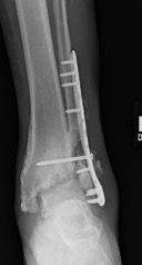
The patient is initially lost to follow up but shows back up in your office eight months later with an untreated draining sinus over the lateral malleolus. He says this wound has opened and closed over the past eight months. New radiographs (shown below) are significant for osteomyelitis of not only the fibula but likely the distal tibia and talus as well. There is also complete destruction of the ankle joint.

This patient has a chronic wound infection, retained hardware and probable osteomyelitis. How do we triage this patient for optimal limb salvage? What are our options? What does the literature tell us about infected hardware?
First of all, in cases of infected hardware and osteomyelitis of the ankle joint, amputation is always an option. It is important to have an honest conversation with the patient about the severity of the diagnosis and what the postoperative course of limb salvage would look like. After your team and the patient are on the same page about goals and expectations, it is important to evaluate whether you are dealing with an acute infection following ORIF, a chronic infection with a stabilized, healed fracture or the worst case scenario in which the fracture has not healed yet and an infection is present. The case I have presented above appears to be a chronic, infected nonunion.
Infected hardware in an already healed fracture setting may be easier to deal with since the joint has mechanical stability. One should remove the hardware and obtain bone biopsies and cultures after multiple surgical debridements. Infectious disease consults are essential as the patient will likely need long-term antibiotics based on culture and biopsy results. If there is extensive bone loss after debridement, stabilization with external fixation and subsequent bone transport/ lengthening versus grafting may be indicated.1
The literature is scarce on this topic but a 2013 study by Ovaska and colleagues looked at predictors of poor outcomes following deep infection after internal fixation of ankle fractures.2 They looked at 1,923 consecutive ankle fractures and identified 97 with deep infections needing surgical debridement. They found that independent factors for increased risk of failure were smoking, malreduction and hardware removal from ununited fracture. They recommend that surgeons only remove the hardware after confirming fracture union.
Prior to any definitive fixation or staged fusion, one should obtain and analyze repeat bone cultures. Fixation depends on bone quality, bone volume and patient goals. In the X-rays presented with the case, there is complete ankle joint destruction present. After extensive surgical debridement, six weeks of IV antibiotics and repeat negative bone cultures, I performed an ankle fusion.
These are complicated cases that should be reserved for surgeons at university or high-volume centers, where a thorough team approach and patient-centric model are implemented. Please email or post any questions or comments below.
Dr. Pirozzi is a Fellow of the American College of Foot and Ankle Surgeons (ACFAS) and serves as Vice President for ACFAS Region 2. She is in private practice in Phoenix.
References
- Runguang L, Guozheng A, Chaojie C, Yirong C, Geohong R. Bone transport for treatment of traumatic composite tibial bone and soft tissue defects: any specific needs besides the Ilizarov technique? Biomech Res Int. 2020. Available at https://www.ncbi.nlm.nih.gov/pmc/articles/PMC7060447/ . Published February 24, 2020. Accessed March 31, 2020.
- Ovaska MT, Makinen TJ, Madanat R, Vahlberg T, Hirvensalo E, Lindahl J. Predictors of poor outcomes following deep infection after internal fixation of ankle fracture. Injury. 2013;44(7):1002-1006.
Additional References
- Klouche S, El-Masri F, Graff W, Mamoudy P. Arthrodesis with internal fixation of the infected ankle. J Foot Ankle Surg. 2011;50(1):25-30
- Papineau LJ, Alfageme A, Dalcourt JP, Pilon L. Chronic osteomyelitis: open excision and grafting after saucerization (author’s translation). Int Orthop. 1979;3(3):165–17
- Kollig E, Esenwein SA, Muhr G, Kutscha-Lissberg F. Fusion of the septic ankle: experience with 15 cases using hybrid external fixation. J Trauma. 2003;55(4):685–691.
- Scranton PE Jr. Use of internal compression in arthrodesis of the ankle. J Bone Joint Surg Am. 1985;67(4):550–555.


