

What You Should Know About Potentially Malignant Wounds
Even if a wound appears to be benign, one must obviously be vigilant against the possibility of malignancy. These expert panelists discuss identifying malignant wounds, taking biopsies and when one might consider an amputation.
Q: What clinical insights lead you to suspect that a lower extremity wound may have an underlying malignancy?
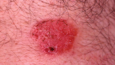
A: M. Joel Morse, DPM, suspects malignancy if a wound does not look like it should. For example, if a neuropathic wound does not behave like it should with offloading, one should suspect melanoma. If a wound shows inadequate healing and a different treatment does not improve the condition, he suggests obtaining a biopsy.
“Do not be tentative about doing a biopsy,” maintains Dr. Morse. “Many times a melanoma or a squamous cell carcinoma does not ‘look’ like a melanoma or squamous cell carcinoma.”
Bradley Bakotic, DPM, DO, says the most important clue is the disconcordance between the development of the lesion and the clinical setting in which it occurs. In other words, he says an “ischemic” ulceration should not arise in well vascularized patients and “stasis” ulcerations are not expected in patients with normal venous return. Prior to attributing an ulceration to a particular etiology based on its appearance, he advises clinicians to verify that the patient actually has the causative condition.
“It is an undisputable fact that basal cell carcinomas can look identical to stasis ulcerations, melanomas may closely resemble ischemic ulcers and squamous cell carcinomas may mimic neuropathic ulcerations,” explains Dr. Bakotic. “A unifying concept is that ‘spontaneous’ ulcerations should be considered neoplastic until proven otherwise.”
Larry Goss, DPM, and Sabrina Minhas, DPM, say the signs of a possible malignancy include any lesion that appears suddenly and changes appearance in shape, size, border irregularity and color. They also note the potential danger of any chronic wound that persists over a long period of time and does not respond to treatment or signs of healing. They note that these patients may have squamous cell carcinoma or Marjolin’s ulceration.
Furthermore, Drs. Goss and Minhas say any lesion that feels deep on palpation and feels fixed to underlying tissues may be malignant as could a large solid lesion that does not transilluminate light. They note that pain or a history of trauma may not be reliable since some malignancies are painless until later stages.
“Appearance of an enlarging mass, although suspicious, is usually not in itself diagnostic for malignancy because the clinical characteristics of malignant lesions are typically varied,” say Dr. Goss and Dr. Minhas. “Physicians must take note of constitutional signs and symptoms, examine and palpate for lymphadenopathy, and take a thorough history and physical to rule out other systemic metastases.”
Both Dr. Goss and Dr. Minhas warn that clinicians sometimes overlook important information like age (some tumors only occur in a certain age group), usual syndromes (i.e. Gardner’s syndrome, Cowden syndrome, Ollier’s syndrome, etc.) and occupational exposure (i.e. radiation, arsenic, etc.).
Q: What are the most common lower extremity malignant wounds that you encounter?
A: Drs. Goss and Minhas categorize the origins of malignant lesions in the foot and ankle in three groups: soft tissue, osseous and distant metastasis. Soft tissue malignancies are more common than osseous counterparts, according to Drs. Goss and Minhas. Malignant soft tissue lesions are derived from ectoderm and include processes from the integument (skin) and glands. They note the common appearance of malignant melanomas (five types: superficial spreading, basal cell, acral lentiginous, lentigo and amelanotic).
All the panelists cite squamous cell carcinoma as common in their facilities with Dr. Bakotic seeing that malignancy daily. Drs. Goss, Bakotic and Minhas also note the common occurrence of basal cell carcinoma.
Less common malignant skin lesions include verrucous carcinoma and Marjolin’s ulceration, concur Drs. Goss and Minhas. They also note that malignant osseous lesions and sarcomas (derived from the mesoderm) are far less common in the lower extremity. Dr. Goss and Dr. Minhas point out that sarcomas that usually present in the foot and ankle include Kaposi’s sarcoma, synovial sarcoma, malignant Schwannoma, osteosarcoma, malignant fibrous histiocytoma, liposarcoma, clear cell sarcoma and, to a lesser extent, fibrosarcomas.
In the pediatric population, they concur that Ewing’s sarcoma and rhabdomyosarcoma are the main etiology. Distant metastasis in the foot and ankle is extremely uncommon. They note that the four most common sites are the lung, bladder, breast and kidney.
Dr. Bakotic notes that since melanomas manufacture abundant brown-black pigment in most cases, physicians are less likely to confuse these lesions with non-neoplastic wounds. However, he says amelanotic melanoma nearly uniformly presents as a non-healing ulceration. Less commonly, he encounters neoplasms such as diffuse large B-cell lymphoma, Merkel cell carcinoma and adnexal carcinoma masquerading as benign ulcerations.
Q: Do you have any insights into how and when to biopsy these wounds?
A: If such wounds are not healing, Dr. Morse takes biopsies. From the start, his treatment plan entails taking biopsies of patients with chronic wounds.
Generally, Dr. Bakotic says for the purpose of histopathologic analysis, one should sample any ulceration that arises spontaneously. For ulcers that appear to occur in concordance with a bona fide causative condition but fail to improve despite appropriate therapy, he says one should sample them after roughly two to three months. Dr. Bakotic warns that failure to sample neoplastic ulcers after two to three months of targeted care could result in establishing causation.
If one encounters superficial wounds that appear to be benign, Drs. Goss and Minhas say one can perform a shave biopsy or a punch biopsy (especially for subungual lesions). They note that physicians can perform fine needle and core biopsies in the office but emphasize getting enough of a specimen for analysis. Physicians may do excisional biopsies for smaller lesions they suspect of being benign whereas incisional biopsies are best for larger lesions that one suspects to be malignant. For any wound suspected of malignancy, they say one should biopsy it as soon as possible to ensure accurate and timely diagnosis, and to facilitate proper care.
Q: When is amputation a consideration?
A: As Dr. Bakotic says, the likelihood of amputation varies widely, depending on the neoplasm’s biology, the affected location and the patient’s level of activity, vascular status and overall health status. Since even the excision of small malignancies of the toes will lead to “monumental problems” with closure, he advises that patients in poor health or those who are inactive will typically fare much better with digital amputation. Younger or largely healthy people are more likely to tolerate plastic procedures, graft placement or healing by secondary intention when it comes to excising carcinomas or in-situ melanomas of the digits, according to Dr. Bakotic. Drs. Goss and Minhas say amputation is a consideration when the patient requires an optimal functional outcome, especially in terms of his or her goals for rehabilitation. For example, they note that large, destructive tumors of the digits may need a distal Symes amputation.
Drs. Goss and Minhas note that aggressive lesions of the forefoot may require either partial ray or transmetatarsal amputation whereas such tumors in the rearfoot usually require a below-the-knee amputation. They concur that most synovial sarcomas and fibrosarcomas usually necessitate amputation as definitive treatment.
In most cases, Dr. Bakotic says thin invasive melanomas of the digits will warrant amputation at the interphalangeal joint and invasive melanomas of greater than 1 mm in thickness will result in amputation at the metatarsophalangeal joint (MPJ). He says amputations proximal to the digits are rarely indicated for tumors of the skin. Dr. Bakotic notes that physicians should reserve proximal amputations for sarcomas of the deep soft tissue that are unlikely to be confused with non-neoplastic ulcerations.
If he finds a malignant wound, Dr. Morse usually consults an oncologist. If an excision is inadequate, he says amputation is the next step. He adds that this requires a certain bedside manner in order to explain the nature of the problem to the patient.
Q: Do you have any insight on nail bed wounds that have a malignant potential?
A: “If there is inadequate healing of a wound, you must biopsy,” emphasizes Dr. Morse. “No matter how many nail bed wounds you have seen, you cannot tell the malignant ones from the benign ones by looking at them. You must biopsy these wounds. If you are concerned about the biopsy, send it to another doctor for a consultation.”
Dr. Bakotic notes the complicating feature in this location is the presence of an overlying nail plate that may or may not be dystrophic. As a result of the anatomy in this location, “wounds” of the nail unit often clinically resemble a subungual hematoma or periungual ecchymosis, according to Dr. Bakotic. Unfortunately, he says such a clinical appearance serves as a diagnostic pitfall and is further complicated by the fact that many of these patients offer a clinical history that includes localized trauma. He estimates that nearly 90 percent of the nail unit melanomas he sees in practice are associated with a false history of “trauma.”
Additionally, Dr. Bakotic notes a little known fact that nail unit malignancies that arise within the matrix (melanoma and some squamous cell carcinomas) can distort nail growth, resulting in a dystrophic nail that may be identical to that created in association with microtrauma or onychomycosis. Since melanoma of the nail unit typically arises within the nail matrix, he says amelanotic lesions are commonly associated with this phenomenon.
For Drs. Goss and Minhas, wounds that begin under the nail and extend outward onto healthy peri-nail skin (Hutchinson’s sign) are a concern due to the possibility of subungual (malignant) melanoma. Amelanotic melanomas of the nail bed often resemble chronic paronychias. Both doctors concur that malignant wounds may form rapidly or may be longstanding nail bed ulcers such as squamous cell ulcers that do not show signs of improvement. They note that such lesions may also appear as ingrown toenails that do not seem to improve no matter what the treatment. While rare, metastases from other primary tumors may develop under the nail as well, according to Drs. Goss and Minhas.
Dr. Bakotic is the Director of the Institute for Podiatric Pathology in Pompano Beach, Fla. He is also affiliated with Dermpath Diagnostics, Ameripath in Pompano Beach, Fla. He is also a Diplomate of the American Board of Pathology.
Dr. Goss is a Fellow of the American College of Foot and Ankle Surgeons. He is the Residency Director of Solis Roxborough Memorial Podiatric Medical & Surgical Residency (PM&S-36) in Philadelphia.
Dr. Minhas is a second-year podiatric surgical resident (PGY-2) at Solis Roxborough Memorial Hospital in Philadelphia.
Dr. Morse is the President of the American Society of Podiatric Dermatology. He is a Fellow of the American College of Foot and Ankle Surgery, and the American College of Foot Ankle Orthopedics and Medicine. Dr. Morse is board certified in foot surgery.
Dr. Karlock is a Fellow of the American College of Foot and Ankle Surgeons, and practices in Austintown, Ohio. He is the Clinical Instructor of the Western Reserve Podiatric Residency Program in Youngstown, Ohio. Dr. Karlock is a member of the Editorial Advisory Board for WOUNDS, a Compendium of Clinical Research and Practice.


