

Orthobiologics: Can They Be Effective For Osteochondral Lesions?
Given the prevalence of hallux abducto valgus deformity, these authors offer a prmer on micro-and macrobiological fundamentals involving the first metatarsophalangeal joint (MPJ) and the impact of joint surface deterioration. They also examine an emergine implant for repair of first MPJ osteochondral lesions. It has been estimated that 209,000 patients undergo surgery for hallux abducto valgus correction each year in the United States.1 The National Center for Health Statistics states that hallux abducto valgus affects 1 percent of the adult population in the U.S.2 Hallux abducto valgus (HAV) deformities have traditionally been classified as ranging from mild to severe. A recent study by Engel correlated the relationship of subchondral bone cyst formation of the first metatarsophalangeal joint (MPJ) and the severity of HAV deformity.3 The intraoperative repair of subchondral defects is imperative during the surgical correction of HAV for articular force distribution during propulsion and to circumvent progression of the osteolytic lesion. Current orthobiological options offer an alternative to the traditional microfracture or marrow stimulating technique (MST). New orthobiological implantable materials have demonstrated evidence of resurfacing the articular surface with hyaline-like soft tissue that adheres to the osteoconductive, bioabsorbable substrate.
Key Insights On Micro- And Macrobiological Concepts
Having a strong understanding of “normal” histology allows for the differentiation of pathological processes. Osteophyte production, lipping of the joint and osteochondral lesions are all indications of a pathological process. The diarthrotic joint is grossly comprised of the soft tissue structures adjacent to and including the bone that provides the foundation of the two (or more) adjacent articular surfaces. The articular surfaces are composed of hyaline cartilage devoid of perichondrum. The hyaline cartilage is comprised of calcified and uncalcified layers. The uncalcified layer is closest to the articular surface and has three distinct zones. The uncalcified layer is anchored to the subchondral bone by the calcified layer of hyaline cartilage. Surrounding the articular surface are plicated folds of synovial membrane that smooth out as their distance from the articular surface increases. Ligaments, tendons (rare) and a dense fibrous capsule support the synovial membrane. The synovial cells filter the adjacent capsular blood supply for water and ions to be delivered into the joint. The synovial cells, which are classified into type A and type B cells, also have other functions based on their respective type. Type A cells are phagocytic and produce hyaluronic acid while the type B cells produce various proteins. Hyaline cartilage is comprised of chondrocytes and surrounding matrix. It does not have a blood supply, lymphatics or enervation. The matrix provided by and surrounding chondrocytes is composed of type II collagen, water and chondroitin sulfate. The combination of water and chondroitin sulfate provides excellent compression strength. Some of the synovial fluid from the joint can diffuse into the outermost layer of the hyaline cartilage. The synovial fluid can then exude upon compression, allowing for instant articular surface lubrication. The pathological process of joint surface deterioration is known to occur via the alteration of the dual anabolic and catabolic function of the chondrocytes. Pathological processes cause the chondrocytes to enzymatically degrade the matrix at a rate faster than their production of the matrix. Degradation of the matrix is caused by activation of catabolic enzymes and decreased production of inhibitors. Mediators of this process are cytokines IL-1 and TNF. Destruction of the articular surface initiates the pathogenesis of the subchondral aberrations. An increase in subchondral bone density may or may not precede a diminished articular surface.
What About The Link Between Subchondral Cysts And HAV?
Subchondral cyst formation in the first MPJ can be attributed to many different etiologies, including trauma and inflammatory disease. Traumatic etiologies can be sub-classified into morbidity of an acute trauma and morbidity of biomechanical/structural malarticulation such as hallux valgus/hallux rigidus leading to osteoarthritis. Inflammatory etiologies include rheumatoid arthritis, seronegative spondyloarthropathies and crystalline arthropathies. Recent evidence supporting concomitant findings of subchondral cyst formation and the severity of HAV infers an increased prevalence of this associated pathology. In a retrospective review, Engel, et al., examined 265 first MPJ of 195 patients (with a mean age of 54.2) for the existence of cartilaginous lesion. The researchers found a statistically significant correlation between the grade of the cartilage damage and the hallux valgus angle.3 One should consider concomitant treatment of cartilaginous injuries during the surgical correction of HAV. Forms of treatment include curettage, microfracture/marrow stimulating technique (MST), decompression osteotomies, autograft/allograft and absorbable synthetic grafts and/or joint arthroplasty with implant or fusion.
Comparing The Marrow Stimulating Technique And The Use Of A Synthetic Implant
The marrow stimulating technique (MST) of reconstituting an articular surface from an articular defect is based on the premise that the resulting blood will form a “soft” callus within the defect and develop into connective tissue (fibrocartilage). At the onset of a simplified model of subchondral microfracture, the blood in the medullary sinus, which includes the stem cells and their bioactive agents, permeates into the defect. If the synovial fluid from the defect does not continually dissipate the blood, this leads to a blood clot, which stabilizes the microfractured surface. Platelets in the clot release platelet-derived growth factor (PDGF) and cytokines, which stimulate the pericytes of the endosteum. The decreased action signals the modification of the stimulated pericytes to transform into chondrocytes. The chondrocytes form the provisional (soft) callus composed of fibrocartilage. The fibrocartilage has mostly type I collagen with some type II collagen and chondroitin sulfate. A different coefficient of friction exists in the comparison of hyaline and fibrocartilage wherein fibrocartilage is less desirable as an articular surface. A recent study of mature Spanish goat knee joints demonstrates the MST may not even result in the formation of the fibrocartilage plug as previously believed.4 However, the same study used OsteoCure (Nexa Orthopedics), a microbiological synthetic implant, to replace the iatrogenic defects. After various stages of healing, the goats were sacrificed and researchers performed cross-sectional histologic analysis of the involved articular surfaces, which were stained for collagen type. Researchers used three different stains to confirm the presence of hyaline-like/hyaline cartilage. The Safranin-O/Fast Green and Toluidine Blue (TMB) stains highlight the cartilage tissue and columnar architecture one normally sees with this type of tissue. The Safranin-O stains proteoglycans and the red/orange color confirms the presence of a hyaline repair cartilage. The Toluidine Blue (TMB) staining confirms the presence of Type II collagen in the defect and the surrounding intact tissue. One will find Type II collagen in hyaline cartilage whereas clinicians will find Type I collagen in fibrocartilage. At six months, a significant amount of cartilage resurfacing had taken place and was still ongoing. Histologically, the trichrome stain stains bone green and cartilage pink. After six months, there is much new bone in the subchondral area and, most importantly, the subchondral bone has bridged to form a support for the overlaying cartilage. There was some implant material left in the subchondral bone. However, this is to be expected since the product is designed to be resorbed in a six- to 12-month time frame. Across the articular cartilage layer, hyaline-like tissue formed in the site formerly occupied by the implant. The tissue appeared to be well integrated with no margin line. Staining was consistent with the surrounding intact tissue. If this were fibrocartilage, the stain would appear different in this area. At 12 months, the defects have continued to heal more completely. The original location of the surgically caused defect is much more difficult to define. Repaired tissue has an even, thick and fairly smooth consistency. Histology sections stained with TMB stains show sulfated glycosaminoglycans (GAGs), a major component of normal articular cartilage, in deep blue/purple. Histologically, the OsteoCure plug has been completely replaced with bone and hyaline-like tissue spans the entire surface of the treated area. The findings noted type II collagen within the confluent articular surface and an osseous substrate anchoring and producing hyaline-like tissue where the OsteoCure implant was incorporated and replaced by completely organic structures.
Can A Synthetic Implant Facilitate Osteochondral Repair?
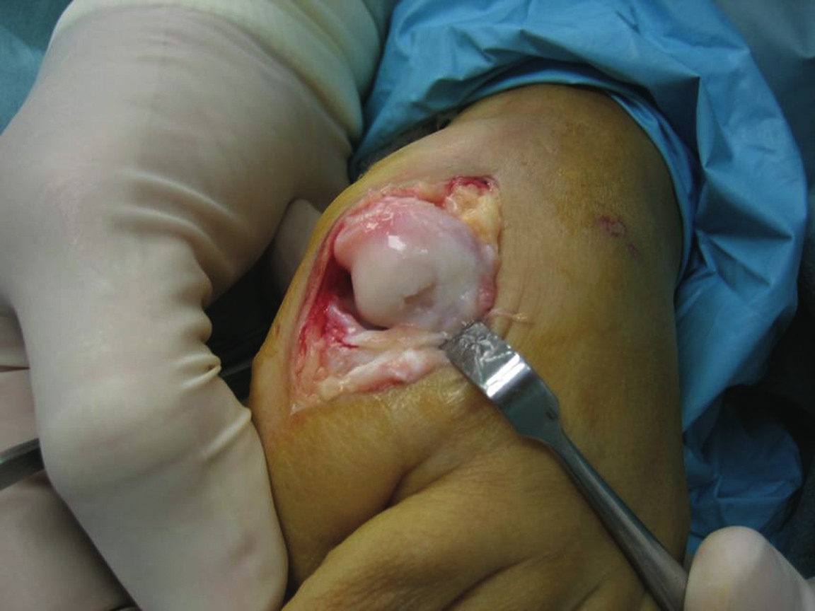
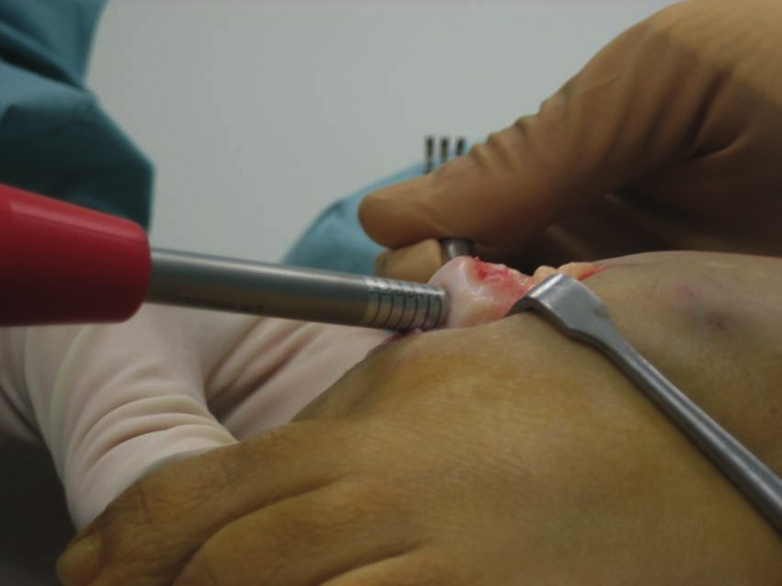
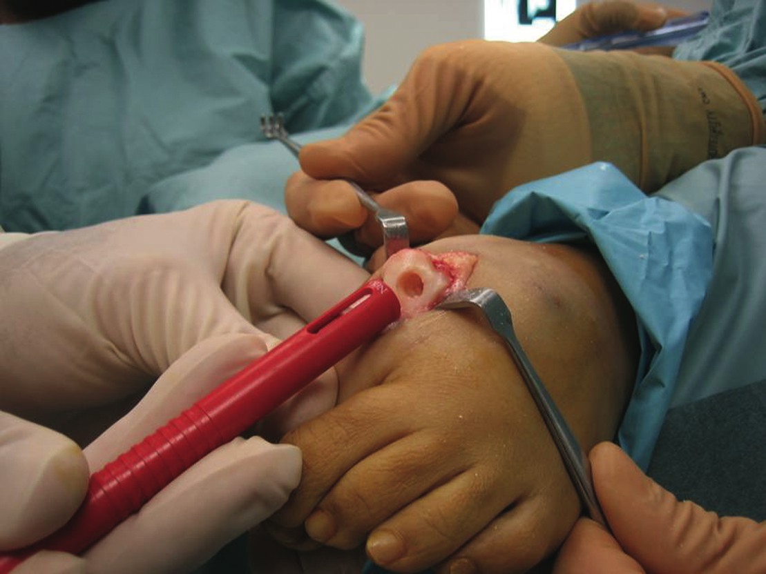
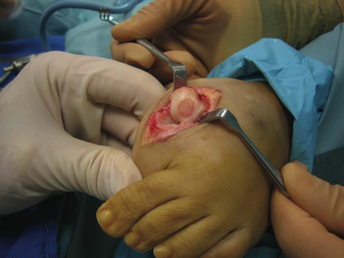
We recently initiated a study of a cohort of 14 patients with hallux valgus and/or hallux rigidus with an osteochondral defect (OCD) who have undergone osteochondral repair of the first metatarsal with the OsteoCure, a synthetic implantable material composed of porous PGA, porous PLG, surfactant and calcium sulfate. All the patients were female and the average age was 51.8. All study participants underwent pre-op X-ray evaluation and range of motion measurements (dorsiflexion/plantarflexion at the first MPJ). We also utilized preoperative magnetic resonance imaging (MRI) evaluations and the Lepow/Chang (L/C) classification of osteochondral lesions (see “A Guide To Classifying Osteochondral Lesions” below). Patients also reported their pain level via a modified 1-10 Visual Analog Pain Scale (VAS). In this study, we employed variable osteotomies and types of fixation to correct the deformities we identified through radiographic and intraoperative assessment. The patients did not have any systemic inflammatory diseases and did not have concurrent untreated osteoporosis or osteomalacia. For the purposes of the study, we excluded patients with bone tumors in the adjacent area of the osteochondral lesion. Our goals with the study were to reduce pain, increase range of motion and develop a functional joint. We also wanted to develop a clear understanding of any evidence displaying the reconstitution of hyaline cartilage at the defect site and complete replacement of the implantable synthetic material with osseous substrate.
Closer Look At The Preliminary Study Results
Early postoperative results with our study reveal excellent results. Postoperative range of motion at the affected first MPJ is increased significantly as compared to the preoperative range of motion. After the immediate postoperative pain and swelling subside, the joint pain is either markedly reduced or is absent in each of the two (or more) adjacent articular surfaces. All patients have returned to impact aerobic exercise, often at an increased level. We expect to see postoperative MRI changes at 24 months that will be consistent with chondral and subchondral repair, and replacement. We will be reporting on the ongoing results of this study in future publications and scientific posters.
Final Notes
The possibility of converting the articular surface of a joint with localized osteochondral pathology to a functional, structural analogue similar to the adjacent non-pathologic bone encourages our given theory. Facilitating the distribution of articular forces across the most anatomically available smooth joint surface or reduced abnormal force distribution could lead to further progression of subchondral bone remodeling and reduced foreseeable morbidity of sequela secondary to cystic pathology. Dr. Lepow is an Associate Professor at the Baylor College of Medicine and University of Texas Medical School in Houston. He is also a Senior Attending Physician at the Houston Podiatric Foundation in Houston. Dr. Smith is an Attending Physician at the Houston Podiatric Foundation. Dr. Sheedy is a Chief Resident at the Houston Podiatric Foundation.
References:
References 1. Coughlin MJ. Hallux valgus: an instructional course lecture. The American Academy of Orthopaedic Surgeons. JBJS (Am) 1996:78-A; 932-66. 2. Graves EJ Detailed Diagnoses and Procedures. National Hospital Discharge Survey 1987. National Center For Health Statistics. Vital Health Statistics 1989: 13(100). 3. Bock P, Kristen KH, Kroner A and Engel A. Hallux valgus and cartilage degeneration of the first metatarsophalangeal joint. JBJS (British Volume) 2004: 86-B(5); 669-673. 4. Sharma B, Fermanian S, Cascio B. Chondral lesion repair in a goat model using an integrated hydrogel scaffold and marrow stimulation. Poster, American Academy of Orthopaedic Surgeons, 2006. Additional References 5. Camasta C. Hallux limitus and hallux rigidus. Clinical examination, radiographic findings, and natural history. Clin Podiatr Med Surg 1996: 13(3); 423-48. 6. Levine B, Kanat IO. Subchondral bone cysts, osteochondritis dissecans, and Legg-Calv-Perthes disease: a correlation and proposal of their possible common etiology and pathologenesis. Journal of Foot Surgery 1988: 27(1);75-9. 7. Roukis T, Weil L Sr., Weil L Jr., Landsman A. Predicting articular erosion in hallux valgus: clinical, radiographic, and intraoperative analysis. JFAS 2005: 44(1);13-21.


