Diagnosis and Management of Gout in the Long-Term Care Setting
INTRODUCTION
Gout is a clinical syndrome caused by tissue (synovial, bursa, cartilage) deposition of monosodium urate (MSU) crystals in a patient with an elevated body pool of uric acid.1 New-onset gouty arthritis is common in the elderly population, and prevalence of gout is on the rise. In a study using a managed care population database, the prevalence of gout and/or hyperuricemia (defined as serum uric acid level > 6.8 mg/dL) in persons over age 75 years increased from 20.55 to 41.28 cases per 1000 enrollees over a 10-year period.2
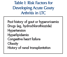
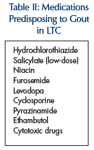
 This seems to be related to increasing lifespan, and thus the age-related diseases such as hypertension and effects of associated treatment as with diuretics (see Tables I and II). Under age 65 years, men are affected four times the number of women.2 Due to loss of the uricosuric effect of estrogen with increasing age, this ratio is reduced to 3:1 after age 65 years. The majority of patients who develop gout have been hyperuricemic for about two decades. However, the majority of individuals with hyperuricemia never develop gout. Most patients with idiopathic gout have a defect (possibly inherited) leading to underexcretion of uric acid, even when the renal function is otherwise normal.1 Diuretic therapy, renal insufficiency, hypertension, and hypertriglyceridemia can be other contributing factors. It has been estimated that at least half of patients who present with an initial attack of gout are taking a diuretic. Approximately 10% of patients
  with gout have increased uric acid production, resulting from excessive purine ingestion, alcohol abuse, genetic defect of purine synthesis, or increased nucleic acid turnover as in myeloproliferative and lymphoproliferative disorders. Gout in the elderly differs from classical gout found in middle-aged men and middle-aged women in several respects: it has a more equal gender distribution, frequent polyarticular presentation with involvement of the joints of the upper extremities, fewer acute gouty episodes, a more indolent chronic clinical course, and an increased incidence of tophi.4
Gout has four distinct stages:
1. Asymptomatic hyperuricemia
2. Acute gouty arthritis
3. Intercritical gout
4. Chronic tophaceous gout
CLINICAL FEATURES
Acute Gouty Arthritis
In an acute presentation, patients will notice severe pain, redness, swelling, and warmth in one or more joints. Severe tenderness will be noted in involved joints. Symptoms worsen within first 24 hours. Joint involvement (in order of decreasing frequency) includes the metatarsophalangeal joint (podagra), the instep/forefoot, the ankle, the knee, the wrist, and the fingers.5 In elderly women, an initial presentation may be acute arthritis of fingers, having inflamed Heberden’s and Bouchard’s nodes.3 Untreated acute gout usually resolves within 1-3 weeks.
Intercritical Gout
This is the period between two attacks of gout. Approximately 60% of patients have a second attack within the first year, and 78% have a second attack within 2 years. Only 7% of patients do not have a recurrence within a 10-year period.6
Chronic Tophaceous Gout
Tophaceous disease is more likely to occur in patients with the following: a polyarticular presentation, a serum urate level higher than 9.0 mg per dL, and a younger age at disease onset (ie, 40.5 years or younger).7 The rate of urate deposition and, consequently, the rate of tophi formation, correlate with the duration and severity of hyperuricemia.6 The most common sites include the joints of the hands and feet. The helix of the ear, the olecranon bursa, and the Achilles tendon are classic, though less common, locations for tophi. Additionally, urate deposition in kidneys could lead to nephrolithiasis.
DIAGNOSIS
Routine Laboratory Data
Consider obtaining complete blood count, basic metabolic panel, erythrocyte sedimentation rate, and serum uric acid level routinely in patients with acute gouty arthritis. Serum urate concentration may reduce during an acute attack. Thus, a normal urate concentration at this point does not rule out a diagnosis of gout.8 If a rheumatoid factor is being obtained due to concern of rheumatoid arthritis as a differential diagnosis, it is important to remember that a rheumatoid factor test is positive in about 30% of patients with tophaceous gout, a finding that relates to the coating of crystals by IgG.3,9 In patients who are candidates for urate-lowering therapy, determination of 24-hour urine uric acid and creatinine excretion is essential to identify the most appropriate urate-lowering medication and to check for significant preexisting renal insufficiency.
Joint Aspiration and Synovial Fluid Analysis
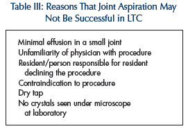
Identifying urate crystals in fluid aspirated from an affected joint is the only definitive way to diagnose gout. Confirmation of the presence of MSU crystals in a patient with signs of acute joint inflammation as local erythema, warmth and tenderness is imperative so that patients with coincidental hyperuricemia and osteoarthritis are not incorrectly diagnosed with gout and unnecessarily treated with allopurinol. It is critical if septic arthritis is being considered as a differential diagnosis of acute gout. MSU crystals are  needle-shaped and negatively birefringent on simple polarized light microscopy. In an acutely inflamed joint, these crystals are seen in polymorphonuclear cells. In practice, however, joint aspiration attempts may not always be feasible in the long-term care setting for a variety of reasons (Table III). The American College of Rheumatology criteria,11 listed below, are helpful in such scenarios to help make the diagnosis. Six of 12 criteria are to be met to make such diagnosis:
1. More than one attack of acute arthritis
2. Maximum inflammation developed within 1 day
3. Attack of monoarthritis
4. Redness over joints
5. Painful or swollen first metatarsophalangeal joint
6. Unilateral attack on first metatarsophalangeal joint
7. Unilateral attack on tarsal joint
8. Tophus (proved or suspected)
9. Hyperuricemia
10. Asymmetric swelling within a joint on radiograph
11. Subcortical cysts without erosions on radiograph
12. Joint fluid culture negative for organisms during attack
Joint fluid accumulations due to acute or chronic gout are nearly always inflammatory in nature, with leukocyte counts between 2000 and 75,000 cells/microliter. Thus, it is important to remember about the leukocyte count overlap between gout and infection. In a patient without previous documentation of gout, aspiration of tophus may help establish diagnosis. However, tophi are often diagnosed clinically due to their location, and an aspiration may thus be avoided since it does carry a risk of infection and subsequent protracted healing. If aspiration is attempted, after infiltrating the area with local anesthetic, a 22-25 gauge needle is used to obtain sample. Since tophi tend to be acellular, MSU crystals in aspirate of tophi may not be associated with polymorphonuclear leukocytes.12
Radiological Evidence
Radiography of a joint affected by gout may show joint space narrowing and destruction of joint surface.7 Tophi may appear as soft-tissue swellings. Additionally, bony erosions with overhanging edges or erosions with sclerotic borders, called punched-out lesions, may be seen on x-rays. Magnetic resonance imaging may show bone “edema,” soft tissue pannus, and swelling.13
THERAPY
The goals of therapy in the management of gout are to alleviate pain and inflammation in acute gouty arthritis, prevent recurrences of acute attacks, and prevent or reverse the complications of urate deposition in kidneys and other involved sites.
Asymptomatic Hyperuricemia
The potential risk of treating asymptomatic hyperuricemia with urate-lowering drugs outweighs benefits since the reaction to medications can be severe and, rarely, even fatal. Asymptomatic hyperuricemia should not be treated per se unless uric acid level is over 13 mg/dL for males and 10 mg/dL for females or in setting of tumor lysis syndrome.1 Otherwise, associated factors such as stopping contributing medications and treating comorbidities such as hyperlipidemia and hypertension should be addressed. Attempts may be made to cut down on such purine-rich foods as organ meats (eg, brain, kidney, liver, pancreas/sweetbreads), red meat, scallops, sardines, beans, lentils, spinach, and mushrooms. In addition, an attempt should be made to stop medications contributing to hyperuricemia such as diuretics, low-dose aspirin, or niacin, among others (Table II), on a case-by-case basis, considering a patient’s overall medical condition.
Acute Gouty Arthritis
Nonsteroidal anti-inflammatory drugs (NSAIDs).
In acute gout, anti-inflammatory dosages of NSAIDs should be given immediately after the onset of symptoms or at the time of diagnosis, and continued for 24 hours after complete resolution of the acute attack, then tapered quickly over 2-3 days (Table IV). Starting NSAID treatment immediately at anti-inflammatory doses is more important than which specific agent is employed. In more than 90% of patients, complete resolution of the attack occurs within 5-8 days of initiation of therapy.14 However, use of NSAIDs in the long-term care setting is limited by the fact that their side effects are more pronounced in elderly patients.15 NSAIDs should be avoided in elderly with creatinine clearance < 50 mL per minute, history of peptic ulcer disease, hepatic dysfunction, poorly controlled congestive heart failure, being on anticoagulation therapy,5 or in postoperative periods. NSAIDs have no effect on serum uric acid levels. While COX-2 inhibitors have not been widely studied in acute gout, they could be considered in some patients at risk for gastrointestinal adverse events.
Corticosteroids.
Systemic steroids should be used only after joint sepsis has been ruled out. Since elderly LTC residents are likely to be more prone to side effects of anti-inflammatory doses of NSAIDs, corticosteroids can be used instead to treat acute flare-ups of gout. Systemic complications of corticosteroids such as osteoporosis, cataracts, and peptic ulcer disease are uncommon if used for short term only.16 However, monitoring blood glucose is important while a patient is receiving corticosteroids. In addition to oral prednisone (Table IV), a single dose of intramuscular triamcinolone acetonide may be used in acute gout. Intra-articular steroid injection is also a treatment option once joint sepsis has been ruled out and only one or two joints are involved.

Adrenocorticotropic hormone (ACTH).
ACTH has not been shown to be any more effective than systemic corticosteroids in management of acute gout and results in more rebound attacks.17 Its use requires intact pituitary-adrenal axis. Side effects include stimulation of release of adrenal androgens and mineralocorticoid causing volume overload.4 It should be given in hospital setting only.
Colchicine.
Colchicine is an antimitotic drug derived from the roots of the herb Colchicum autumnale. Once a drug of choice for acute gout, colchicine has fallen out of favor, especially in the elderly due to its low therapeutic index. Colchicine should be used in the LTC setting only if a patient has contraindications to NSAIDs and steroids. Lower doses of colchicine are somewhat effective though less toxic than a traditional regimen. Colchicine can be given in doses of 0.5 mg three times a day.18 Also, at times, a combination of steroids and colchicine has been used in acute gout. Nearly every patient after 1 day of treatment with colchicine has been shown to develop diarrhea, and often even before the pain is completely resolved. This could lead to dehydration in elderly LTC residents. Adverse effects may occur even with low doses of colchicines (< 2 mg) in the elderly.19 Renal, hepatic, and myocardial impairment and presence of cardiac arrhythmias can enhance the risk of colchicine toxicity.3 Deaths have been reported in patients in whom high doses of intravenous colchicine were used in acute gout.20 The risk of death is higher if a patient is already taking oral colchicine.
Oral narcotics.
Gout remains one of most painful conditions known in elderly. Oral narcotics are frequently needed for adequate pain control. If there is a large joint effusion, aspiration of a large amount may reduce the pain.
When should a physician hospitalize a patient? Patients with acute arthritis should be hospitalized if the clinician suspects septic arthritis or, in rare cases, to achieve better pain control in acute gout with parenteral narcotics.
Intercritical and Chronic Tophaceous Gout
It is during this intercritical phase that the physician should focus on secondary causes of hyperuricemia. Medications should be assessed to identify those that may aggravate the patient's condition (eg, diuretics, low-dose aspirin).5 Attempts to change diet to purine-free are often challenging in the LTC setting, and even when successfully done, are only moderately effective in lowering serum uric acid levels over a long period of time.11
Urate-lowering therapy should be initiated in patients who have frequent attacks of acute gout, tophi, uric acid stones, or severe erosive arthritis. Initiate treatment 2 weeks after the acute attack has subsided, unless kidneys are at risk because of unusual uric acid load. Urate-lowering treatment, if started during an acute attack, can lead to delayed response or rebound exacerbation of gout. A retrospective cross-sectional study has shown that maintaining the uric acid level between 4.6 and 6.6 mg/dL prevents recurrences of gout.21 However, a level lower than 5 mg/dL may be required for resorption of tophi.22 Usually this treatment is lifelong.
Uricosuric agents.
Use uricosuric agents when a patient is a hypoexcretor of uric acid (< 800 mg/24 hr on unrestricted diet or < 600 mg/24 hr while on purine-free diet), relatively young (< age 60), has a creatinine clearance over 60 mL per minute, and has no history of tophi or nephrolithiasis. These drugs enhance in urinary elimination of urate by inhibiting proximal tubular reabsorption of filtered and secreted urate.1 This action is inhibited by low-dose salicylates and may account for a significant number of “treatment failures.”
Probenecid.
Probenecid is the most commonly used uricosuric agent. It should be started at 250 mg BID, with very gradual increase with adequate hydration, and increased by 500 mg at monthly intervals, until the uric acid is lowered to 6 mg/dL (maximum dose = 2-3 g/d). The initial side effects of probenecid include possible precipitation of an acute gouty attack and renal calculi. Other common side effects include rash and gastrointestinal problems.5
Other uricosuric agents.
High-dose salicylates, micronized fenofibrate, sulfinpyrazone, and losartan are other uricosuric agents. Consider using fenofibrate in patients with gout and hyperlipidemia since it can lower serum uric acid levels by 20-35%,23 by decreasing renal tubular reabsorption of urate, thus increasing its renal excretion. Sulfinpyrazone is preferred by some physicians because of its added antiplatelet effects. Therapy is initiated at a dosage of 50 mg 3 times a day, which is gradually increased until the serum urate level is lowered.5 Losartan can be used in treating hypertension in patients with gout who need urate-lowering treatment, due to its uricosuric properties.24
Xanthine oxidase inhibitors.
Xanthine oxidase inhibitors are agents of choice for patients with urate overproduction (urate excretion > 800 mg/24 hr), creatinine clearance < 60 mL/min, tophaceous deposits, nephrolithiasis, and those in whom uricosuric agents were ineffective or cannot be used, as when a patient needs the cardioprotective effect of aspirin.1,5
Allopurinol.
Allopurinol is the most commonly used xanthine oxidase inhibitor. Starting dose of allopurinol in the elderly patient is 50-100 mg on alternate days,4 which is increased cautiously in increments of 50-100 mg per day every 2 weeks until the patient's urate level is less than 6 mg/dL. The maximum dosage of allopurinol should be determined by the creatinine clearance. Frequently, in the LTC setting, 100 or 200 mg of allopurinol in a single daily morning dose will suffice. Up to 5% of patients are unable to tolerate allopurinol, most commonly because of rash. Desensitization is indicated under supervision of a rheumatologist in patients who need allopurinol but have experienced mild to moderate rash. However, rarely, a severe rash accompanied by vasculitis, hepatitis, and renal insufficiency may occur and carries a 20% mortality risk.1 Allopurinol toxicity usually occurs within the first month of initiating therapy, and is more likely if the patient has been on hydrochlorothiazide therapy.
Oxypurinol.
Patients who have not tolerated allopurinol but need a urate-lowering therapy need to be referred to rheumatologists to be tried on oxypurinol. Oxypurinol is the major active metabolite of allopurinol and is available in the United States for use on a compassionate basis only.5
Tumor necrosis factor (TNF) inhibitors.
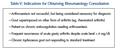
In selected cases, treatment with TNF-alpha inhibitors such as etanercept25 or infliximab26 might be an option in patients with chronic tophaceous gout who do not respond to conventional treatment. Referral to a rheumatologist is recommended in such patients (Table V).

GOUT PROPHYLAXIS
There is a risk of acute gouty flare up when urate-lowering therapy is initiated. Hence, it is common practice among rheumatologists to start prophylactic low-dose NSAIDs or colchicine (from 0.6 mg to 1.2 mg) at the same time that urate-lowering drug therapy is initiated.27 This is continued for at least 6 months after serum urate level has returned to normal. Urate-lowering agent should not be stopped during recurrence of an acute attack if patient has already been taking it.
SUMMARY
There are 10 points to remember in management of gout and hyperuricemia in the LTC setting:
1. Do not treat asymptomatic hyperuricemia.
2. Acute arthritis plus hyperuricemia does not equal gout.
3. At least half of patients who present with an initial attack of gout take a diuretic or other medications predisposing to hyperuricemia or gout.
4. Joint aspiration and synovial fluid analysis is the gold standard in diagnosing gout. If it is not possible or successful in the LTC setting, use the American College of Rheumatology criteria to diagnose gout.
5. Efforts to change diet to purine-free in the LTC setting are challenging and of limited benefit over a long period of time.
6. Prefer short-term use of corticosteroids while avoiding use of colchicine and NSAIDs in managing acute gouty arthritis in elderly residents.
7. Hospitalize any patient with acute arthritis when septic arthritis is being considered as a differential diagnosis.
8. Do not start a urate-lowering agent during an acute attack of gout; however, do not stop one if the patient is already taking it and experiences a recurrence.
9. The starting dose for allopurinol is 50-100 mg every other day, which is increased cautiously every 2 weeks. Usual maximum dose in an LTC resident would be 100-200 mg once a day.
10. Know when to obtain consultation with a rheumatologist.
The author reports no relevant financial relationships.


