An In vitro Comparison of Two Silver-containing Antimicrobial Wound Dressings
Abstract
Preclinical studies have shown that release of silver by different wound dressings varies. The purpose of this in vitro study was to compare the antimicrobial activity of silver alginate (SA) and silver carboxymethylcellulose (SCM) dressings.
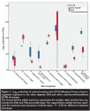
Antimicrobial activity was tested using nine bacterial strains with log10 reduction and corrected zone of inhibition (CZOI) assays. Antimicrobial effect was visualized using confocal microscopy (CLSM). Log10 reduction was comparable between both dressings for Staphylococcus aureus NCIMB 9518, Candida albicans ATCC 90028, Finegoldia magna NCTC 11804T, and Pseudomonas aeruginosa NCTC 10662. Log10 reduction was higher for SCM than SA dressing-exposed Escherichia coli (P = 0.035) and P. aeruginosa ATCC 15692 (P = 0.032), and lower for SCM than SA dressing-exposed Streptococcus pyogenes (P = 0.007), Peptoniphilus asaccharolyticus (P = 0.045), and S. aureus NCTC 8325 (P = 0.012). Both dressings were equivalent against four strains (5 to 8 days’ activity) in the CZOI assay. SA dressing silver activity lasted >24 hours longer than SCM activity when exposed to C. albicans (9 days’ activity), E. coli (7 days’ activity), F. magna (5 days’ activity), and P. asaccharolyticus (5 days’ activity), whereas the SMC exhibited greater persistence against S. pyogenes (13 days’ activity). CLSM showed complete kill of S. aureus after 20 hours for both dressings. The results of this study confirm the broad-spectrum, in vitro activity of some dressings containing ionic silver. The in vitro antimicrobial efficacy of both wound dressings was comparable, but clinical studies comparing the efficacy and effectiveness of silver-containing dressings to nonionic silver-containing dressings are needed.
Potential Conflicts of Interest: This research was funded by a Knowledge Transfer Partnership Award and Dr. Hooper was the KTP associate. KTP funding had been awarded jointly to Advanced Medical Solutions Ltd, Winsford, Cheshire, UK and Cardiff University (Dr. Williams, Prof. Thomas, and Dr. Hill). Dr. Percival was Director of Research and Innovation, Advanced Medical Solutions, Winsford, UK.
Introduction
Chronic wounds affect a high percentage of the worldwide population and their prevalence is increasing. Underlying medical conditions such as obesity and diabetes can induce chronic wound formation and subsequent delayed healing. These wounds generally are thought to exist in a state of continuous inflammation,1 causing them to be considered out of balance, typified by prevention or delay of a proper wound healing response.
Micro-organisms within wounds are known to affect wound balance. Chronic wounds harbor multiple species of micro-organisms that, when combined with underlying patient pathologies, increase the risk of wound infection, which may lead to systemic infection.2 Wound dressings impregnated with topical antimicrobials such as ionic silver are routinely used in the in vivo management of at-risk and infected chronic wounds; clinical studies have documented good clinical patient outcomes.3-6 One important criterion for silver-impregnated wound dressing performance is demonstrated antimicrobial activity that can be sustained over the time of its use against a diverse range of micro-organisms.7 A controlled clinical study3 compared the use of a silver alginate/carboxymethylcellulose (SACMC) dressing with a control nonsilver calcium alginate fiber dressing in the management of 36 patients with venous and pressure ulcers. After 4 weeks of treatment, wounds in the SACMC dressing group showed a statistically significant (P = 0.017) improvement in healing as indicated by reduction in the surface area of the wound compared to the control. Furthermore, in a study6 of 20 patients using cerium nitrate-silver sulphadiazine as a topical burn dressing, a low incidence of actual wound infection was noted, even though bacterial colonization of at-risk burn wounds progressively increased with time after burning.
 A multicenter, randomized, controlled clinical trial study8 of a new silver-releasing dressing (n = 51) was conducted using a nonsilver-releasing control dressing (n = 48) in patients with venous leg ulcers showing signs of inflammation. At the end of the 4-week follow-up period, a significant (P = 0.023) reduction in wound area occurred in the silver group compared with the control group. Importantly, a recent study9 based on measuring corrected zones of inhibition (CZOI) showed the antimicrobial activity of SA against micro-organisms frequently associated with colonization and infection in wounds, including multidrug-resistant (MDR) Pseudomonas aeruginosa, methicillin-resistant Staphylococcus aureus (MRSA), and vancomycin-resistant enterococci (VRE).
Because a wound dressing may be used on a specific patient wound for several days, it is important to know whether the release and efficacy of the antimicrobials contained therein can be maintained over the time period.
The aim of this study was to compare the in vitro antimicrobial activity of two silver-containing wound dressings: RESTORE Silver Alginate (SA) (Hollister Wound Care, Libertyville, IL) and a silver carboxymethylcellulose (SCM) dressing (AQUACEL® Ag Hydrofiber® Dressing; ConvaTec UK, Uxbridge, UK), measuring their antimicrobial performance utilizing a number of antimicrobial efficacy assays.
Methods and Procedures
Dressings. The study test dressings, SA and SCM, have been marketed for many years. Both dressings are highly absorbent and developed to provide a sustained release of silver ions into the wound environment, thereby perpetuating an antimicrobial effect against a wide range of organisms. SA dressings contain silver sodium hydrogen zirconium phosphate and consist of a complex of calcium and ionic silver for effective antimicrobial action and odor reduction. SCM dressings incorporate Hydrofiber® technology and are composed of sodium carboxymethylcellulose and silver carboxymethylcellulose. These versatile, primary dressings are indicated for use on moderately and highly exuding chronic and acute wounds. Results of an in vitro log10 reduction study10 have indicated that this SCM dressing provides rapid and sustained antimicrobial activity for up to 7 days and is effective against P. aeruginosa and S. aureus over this time period.
 Micro-organisms. A total of nine microbial species/strains associated with wound colonization, including both Gram-positive and Gram-negative bacteria as well as yeast, were used to assess the antimicrobial activity of the dressings (see Figure 1). All aerobic species (P. aeruginosa, S. aureus, Streptococcus pyogenes, Escherichia coli, and Candida albicans) were subcultured at 24-hour intervals and maintained at 37° C using Mueller-Hinton agar (MHA) and Mueller-Hinton Broth (MHB). Strict anaerobic bacterial strains (Finegoldia magna, Peptoniphilus asaccharolyticus) were cultured on fastidious anaerobe agar (FAA) supplemented with 5% (v/v) defibrinated horse blood (TCS Biosciences Ltd, Buckingham, UK) and in brain-heart infusion (BHI) broth. These anaerobic bacteria were cultured for 48 hours at 36˚ to 37° C in an anaerobic environment (10% v/v CO2, 20% v/v H2, 70% v/v N2). Unless otherwise stated, all media were obtained from Lab M™ (International Diagnostics Group plc, Bury, UK).
Log10 reduction assay to measure antimicrobial efficacy. The ability of the dressings to kill the micro-organisms within a 2-hour exposure time was determined using a log10 reduction assay based on methods described by Cavanagh et al.11 Briefly, colonies from agar cultures were used to inoculate 20 mL of liquid medium, which was incubated for either 24 hours (aerobes) or 48 hours (anaerobes). A portion of this culture (100 µL) was used to inoculate a fresh 20-mL liquid culture that was incubated under the same conditions until the organism was in log-phase growth and contained approximately 108 colony forming units (CFU)/mL, either for 4 to 6 hours (aerobes) or 36 to 48 hours (anaerobes).
Test dressings were aseptically cut into 2-cm2 pieces and saturated with sterile phosphate-buffered saline (PBS) during incubation (1 hour) in the dark at room temperature. An equal number of control dressing pieces were similarly saturated with a neutralization buffer (NB), which inactivated ionic silver and consisted of PBS containing 1% (v/v) polysorbate 20 and 0.1 % (w/v) sodium thioglycolate (Sigma-Aldrich Ltd, Gillingham, UK). Saturated dressings were aseptically transferred, without squeezing, and allowed to drain for 10 seconds before being placed flat in a sterile container. Each dressing then was inoculated with 1 mL of the log-phase culture and incubated in the dark under the appropriate conditions at 37° C for 2 hours. Following incubation, dressings were placed in NB (9 mL) to achieve a 1:10 dilution of the inoculum and vigorously vortexed for 10 seconds to resuspend the microbial cells. The recovered micro-organisms then were serially diluted 10-fold in NB. Each dilution (50 µL) was spirally plated onto appropriate agar media using a Whitley Automatic Spiral Plater (WASP; Don Whitley Scientific Ltd., Shipley, UK), and these agars were incubated either overnight (aerobes) or for 48 hours (anaerobes). Subsequently, the number of colonies was counted and used to calculate the number of CFU from each dressing. The counts generated from the control dressing pieces were used to estimate the initial number of microbial CFU/mL in the original inocula, while the counts from the experimental dressing pieces were used to calculate the surviving number of CFU/mL. Log10 reduction values, representing the antimicrobial effect of the silver in the dressing, then were calculated as the difference between the log10 values of the starting and surviving numbers of micro-organisms.
Day-to-day corrected zone of inhibition assay to assess antimicrobial longevity. The dressings’ ability to inhibit microbial growth over several days was assessed using a day-to-day agar transfer and CZOI assay. A fresh culture (100 µL) of each test micro-organism was spread evenly onto agar, as per the BSAC guidelines12 for disc diffusion testing. Triplicate pieces of the test dressings (2 cm2) were saturated with sterile distilled water and aseptically placed onto the middle of the seeded agar plates. Before agar placement, the dressings were allowed to drain for approximately 10 seconds. The original dressing placement was traced onto the base of the Petri plate to allow correction for any dressing shrinkage over time. Plates were incubated with the dressing, either overnight (aerobic species) or for 48 hours (anaerobes). The dressings then were transferred to a new agar plate, seeded as above, and incubated in the same manner.
The zones of microbial growth inhibition and original dressing widths were measured in cm in two perpendicular directions for each plate. These measurements then were used to calculate the CZOI by subtracting the dressing width from the inhibition zone width. This procedure was repeated for each micro-organism for up to 14 days or for as many days as required for all the dressing pieces to stop producing any zone of inhibition.
Confocal laser scanning microscopy (CLSM) to visualize death of sequestered S. aureus. S. aureus is one of the most commonly isolated chronic wound organisms. S. aureus is of increasing concern because of the relative ease with which it is able to acquire multiple antimicrobial resistance to a broad range of antibiotics (ie, MRSA). Hence, S. aureus was taken as a model wound organism to determine whether cell death could be observed in vitro within an actual wound dressing to show that the dressing is bactericidal and that it sequesters the bacteria within the actual fibers of the dressing.
For this study, S. aureus NCTC 8325 was cultured in a shaking incubator for 5 to 6 hours until mid-log phase growth. Each culture (5 mL) was briefly centrifuged (13,000 g, 1 minute), the media gently aspirated, and the pellet of cells re-suspended in PBS (1.5 mL) containing working concentrations of LIVE/DEAD® BacLight™ bacterial viability dyes (Invitrogen Molecular Probes, Paisley, UK). The stained bacteria (100 µL) were pipetted onto fibers from each dressing, placed on glass slides with a cover slip, and sealed with petroleum jelly to prevent them from drying out. Preparations were viewed and analyzed using a Leica TCS SP2 spectral confocal microscope and Leica confocal software (Leica, Heidelberg, Germany). Representative regions of dressing were scanned through their full depth approximately 1 hour after inoculation and again after 20 hours’ incubation at room temperature. Scanning was performed using a x20 objective lens and appropriate scan parameters for simultaneous fluorescence recordings of live cells (Syto 9; green fluorescence; Ex. max. 485 nm; Em. max. 500 nm) and dead cells (propidium iodide; red fluorescence; Ex. max. 536 nm; Em. max. 617 nm). The excitation lasers used for each probe (argon 488 nm and helium neon 543 nm, respectively) were used at their lowest possible power output to minimize potential phototoxic side effects on the micro-organisms. To provide context, individual fibers within the dressing were simultaneously imaged using Nomarski differential interference contrast (DIC) optics. Z-Stacks of optical sections taken at each time point were reconstructed using a maximum intensity projection algorithm and then presented as green/red bacterial overlays upon a greyscale DIC image of the fibers. The comparative proportions of live and dead bacteria in each sample could be estimated by analyzing the relative ratio of representative green/red (LIVE/DEAD®) fluorescent signal intensities (ie, voxel intensities 0 to 255) from within selected regions of interest (ROIs).
Statistical analysis. Box plots for log10 reduction assays were prepared using SPSS software (SPSS Inc, Chicago, IL). For the log10 reduction and CZOI assays, comparison of both dressings was achieved using two-tailed two sample t-tests (Microsoft Excel 2007).
Results
The log10 reduction assay demonstrated noticeable antimicrobial activity with both the SA and SCM dressings (see Figure 1). All species tested exhibited inhibition by the silver-containing dressings, although this was in a strain-dependent manner. Antifungal activity (C. albicans) was similar for both dressings, but overall log10 reduction for the Gram-negative strains E. coli and P. aeruginosa ATCC 15692 was significantly greater with the SCM than the SA dressing (P = 0.034 and P = 0.012, respectively). However, the mean log10 reduction for Gram-positive anaerobic bacteria species P. asaccharolyticus was 0.97 for SA compared to 0.2 in the SCM dressing group (P = 0.045). Similarly, the mean log10 reductions for S. aureus NCTC 8325 and S. pyogenes were 1.36 and 2.36 in the SA and 0.73 and 1.23 when exposed to the SCM dressing (P = 0.012 and P = 0.007, respectively). S. aureus NCIMB 9518 showed limited sensitivity to both silver-containing dressings in the 2-hour exposure time compared with the other micro-organisms studied. Differences between dressings in log10 reduction following exposure to all the other strains tested were not statistically significant.
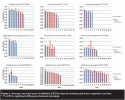
 Results of the CZOI assay showed that antimicrobial activity of the silver-containing dressings lasted from 4 to 13 days (see Figure 2). Both the SA and the SCM dressings exhibited activity against four of the nine strains (two strains each of P. aeruginosa and S. aureus) for a period of 5 to 8 days. A greater persistence of SA than SCM dressing was evident against the two anaerobic species, with activity still detected after 5 days. SA dressing also was observed to retain activity longer than the SCM dressing against C. albicans and E. coli. In contrast, the SCM dressing exhibited a slightly longer-lasting activity against S. pyogenes.
Statistical differences (P <0.05) in the mean CZOI were evident between the dressings for the micro-organisms tested. When both dressings demonstrated activity, the CZOI values for SCM were significantly higher for C. albicans (days 1–2, 4–8), E. coli NCTC 12923 (days 1–3), P. aeruginosa ATCC 15692 (days 2–5), P. aeruginosa NCTC 10662 (days 1–4), S. aureus NCIMB 9518 (days 1–2), S. aureus NCTC 8325 (day 1), and S. pyogenes NCTC 8198T (days 1–4, 7–12). Similarly, the CZOI values for SA were significantly higher for F. magna NCTC 11804T on day 4.
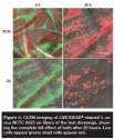
 CLSM fiber imaging from the two dressings inoculated with LIVE/DEAD®-stained S. aureus showed an equivalent antimicrobial effect — namely, complete bacterial kill after 20 hours on both dressings.
Discussion
Controlling the microbial burden within wounds has long been recognized as an important aspect of wound management. Contamination by mixed or single-species populations of micro-organisms can lead to colonization and infection, which can impair wound healing. The use of antimicrobial dressings is advocated to combat this problem,13,14 and in recent years much focus has been given to the potential role of silver-containing dressings in inhibiting the growth of the wound microbiota.11,15-18 The aim of this study was to compare the in vitro antimicrobial activity of two silver-containing wound dressings against a range of common wound micro-organisms. These species were selected based on their prevalence within wounds and pathogenic associations. The methods used, chiefly CZOI and log-reduction assays, were those previously recommended for the analysis of silver-containing dressings.11,19,20
Both the SA and the SCM dressings exhibited antimicrobial activity against all of the tested wound isolates, which included Gram-positive bacteria, Gram-negative bacteria, and fungi. In this and other in vitro studies,21 microbiocidal activity varied between species and strains. The broad-spectrum activity of ionic silver in vitro has been reported before,15,22 although with greater activity against Gram-negative bacteria compared with Gram-positive after 24 hours.15 Similarly, in this study, the log10 reduction assays performed showed that a 2-hour exposure of the silver dressings had a limited effect on one of the two S. aureus strains used. However, during longer exposures (up to 8 days) using the CZOI assay, complete bacterial kill was achieved.
As the activity of the dressings is due, at least in part, to the availability of ionic silver in the dressing, a loss of antimicrobial efficacy over time is to be expected. Previous studies19 often only looked at a single time point (usually 24 hours) for the CZOI testing, but most silver-containing dressings are in place for several days. In this CZOI time course study, both dressings exhibited an antimicrobial effect against all test isolates for a minimum of 4 days. The SA dressing showed similar longevity of antimicrobial efficacy for S. aureus (5 and 8 days) to a previous CZOI time course (6-day) study11 using a similar type of dressing, but the variability between these and current results again suggests this effect is strain-dependent. The ability to retain antimicrobial activity following repeat microbial challenge is important in clinical situations where dressings may remain in place for several days. It was particularly interesting to find that both products tested here demonstrated a persistent effect, when similar studies have reported that some silver-containing foam dressings were not capable of producing CZOIs against wound micro-organisms.11,15,23
The general similarities in antimicrobial activity between the two dressings also was confirmed by the use of LIVE/DEAD® staining and CLSM imaging of inoculated dressing fibers. Observing microbial kill in this way is relatively novel, although a similar approach has been used previously24 to look at the antimicrobial properties of a similar SCM silver-containing hydrofiber dressing. The current study found that the dressing fibers exhibited a degree of autofluorescence visible in the red channel used to detect the dead cells, which precluded using the relative intensity measurements to estimate the proportions of living and dead cells in the samples. However, despite these methodological limitations, it is still a useful technique for visualizing how the bacteria were successfully sequestered and immobilized between the fibers and killed in situ. Importantly, very few clinical studies comparing the efficacy of silver-containing wound dressings to dressings containing no ionic silver have been conducted, being that these studies are needed to interpret the clinical validity of the current in vitro findings.
Limitations and Implications
In vitro studies facilitate dressing material testing by using a large number of challenge organisms and sufficient replicates to ensure statistical validity. Due to limitations in patient numbers, these options are far harder to achieve in in vivo testing. Although in vitro experimentation gives a good indication of how these dressings can work in practice, clinical efficacy and effectiveness require suitable clinical studies, including randomized controlled clinical trials. Because very few clinical studies comparing the efficacy of silver-containing wound dressings to dressings containing no ionic silver have been conducted, these studies are needed to interpret the clinical validity of the current findings.
Conclusion
The purpose of this study was to evaluate and compare the in vitro antimicrobial efficacy of two wound dressings containing ionic silver. Both the SA and the SCM dressing were found to reduce the counts of broad-spectrum of wound micro-organisms 2 hours following exposure. In addition, using the CZOI assay, the dressings were noted to exhibit antimicrobial activity for a minimum of 4 days. These results suggest that both dressings may help manage wound microbial burden, although this needs to be confirmed by further in vitro and clinical studies.
Acknowledgments
The authors acknowledge the support for this research and for Dr. Hooper that was provided by a Knowledge Transfer Partnership (KTP) between Cardiff University and Advanced Medical Solutions, Winsford, UK.
Dr. Hooper is a KTP Associate; Dr. Williams is a Reader in Oral Microbiology; Prof. Thomas is Professor and Honorary Consultant in Oral and Maxillofacial Surgery; and Dr. Hill is a Post-Doctoral Research Fellow; Tissue Engineering and Reparative Dentistry, Cardiff University School of Dentistry, Cardiff, Wales. Dr. Percival is Director of Research and Innovation, Advanced Medical Solutions, Winsford, UK. Please address correspondence to Dr. David Williams, School of Dentistry, Cardiff University Dental Hospital, Heath Park, Cardiff CF14 4XY, Wales; email: WilliamsDD@cardiff.ac.uk.
1. Falanga V. Classification for wound bed preparation and stimulation of chronic wounds. Wound Repair Regen. 2000;8:347–352.
2. Percival SL, Dowd S. The microbiology of wounds. In: Percival SL, Cutting K (eds). Microbiology of Wounds. London, UK: CRC Press;2010:187–218.
3. Beele H, Meuleneire F, Nahuys M, Percival SL. A prospective randomised open label study to evaluate the potential of a new silver alginate/carboxymethylcellulose antimicrobial wound dressing to promote wound healing. Int Wound J. 2010;7(4):262–270.
4. Caruso DM, Foster KN, Hermans MH, Rick C. Aquacel Ag in the management of partial-thickness burns: results of a clinical trial. J Burn Care Rehabil. 2004;25:89–97.
5. Cutting KF. A dedicated follower of fashion? Topical medications and wounds. Br J Nurs. 2001; 10: 9-16.
6. Ross DA, Phipps AJ, Clarke JA. The use of cerium nitrate-silver sulphadiazine as a topical burns dressing. Br J Plast Surg. 1993;46:582–584.
7. Bradford C, Freeman R, Percival SL. In vitro study of sustained antimicrobial activity of a new silver alginate dressing. JACCWS. 2009;1:117–120.
8. Lazareth I, Meaume S, Sigal-Grinberg ML, Combemale P, Le Guyadec T, Zagnoli A, Perrot J-L, Sauvadet A, Bohbot S. The role of a silver-releasing lipido-colloid contact layer in venous leg ulcers presenting inflammatory signs suggesting heavy bacterial colonization: results of a randomized controlled study. WOUNDS. 2008;20:158–166.
9. Percival SL, Thomas J, Linton S, Okel T, Corum L, Slone W. The antimicrobial efficacy of silver on antibiotic-resistant bacteria isolated from burn wounds. Int Wound J. 2011;12:19.
10. Bowler PG, Jones SA, Walker M, Parsons D. Microbicidal properties of a silver-containing Hydrofiber® dressing against a variety of burn wound pathogens. J Burn Care Rehabil. 2004;25:192–196.
11. Cavanagh MH, Burrell RE, Nadworny PL. Evaluating antimicrobial efficacy of new commercially available silver dressings. Int Wound J. 2010;7:394–405.
12. Andrews JM. The development of the BSAC standardized method of disc diffusion testing. J Antimicrob Chemother. 2001;48(S1):29–42.
13. Schultz G, Barillo DJ, Mozingo DW, Chin GA. Wound bed preparation and a brief history of TIME. Int Wound J. 2004;1:19–32.
14. Sibbald RG, Williamson D, Orsted HL, Campbell K, Keast D, Krasner D, Sibbald D. Preparing the wound bed-debridement, bacterial balance, and moisture balance. Ostomy Wound Manage. 2000;46:14–35.
15. Ip M, Lui SL, Poon VK, Lung I, Burd A. Antimicrobial activities of silver dressings: an in vitro comparison. J Med Microbiol. 2006;55:59–63.
16. Kotz P, Fisher J, McCluskey P, Hartwell SD, Dharma H. Use of a new silver barrier dressing, ALLEVYN Ag, in exuding chronic wounds. Int Wound J. 2009;6:186–194.
17. Percival SL, Slone W, Linton S, Okel T, Corum L, Thomas JG. The antimicrobial efficacy of a silver alginate dressing against a broad spectrum of clinically relevant wound isolates. Int Wound J. 2011;8:237–243.
18. Percival SL, Slone W, Linton S, Okel T, Corum L, Thomas JG. Use of flow cytometry to compare the antimicrobial efficacy of silver-containing wound dressings against planktonic Staphylococcus aureus and Pseudomonas aeruginosa. Wound Repair Regen. 2011;19:436–441.
19. Gallant-Behm CL, Yin HQ, Liu SJ, Heggers JP, Langford RE, Olson ME, Hart DA, Burrell RE. Comparison of in vitro disc diffusion and time kill-kinetic assays for the evaluation of antimicrobial wound dressing efficacy. Wound Rep Regen. 2005;13:412–421.
20. Thomas, JG, Slone W, Linton S, Okel T, Corum L, Percival SL. In vitro antimicrobial efficacy of a silver alginate dressing on burn wounds isolates. J Wound Care. 2011;20:124–128.
21. Kim J, Kwon S, Ostler E. Antimicrobial effect of silver-impregnated cellulose: potential for antimicrobial therapy. J Biol Eng. 2009;3:20.
22. Parsons D, Bowler PG, Myles V, Jones S. Silver antimicrobial dressings in wound management: a comparison of antibacterial, physical, and chemical characteristics. WOUNDS. 2005;17:222–231.
23. Newman GR, Walker M, Hobot JA, Bowler PG. Visualisation of bacterial sequestration and bactericidal activity within hydrating Hydrofiber wound dressings. Biomaterials. 2006;27:1129–1139.


