Medical Honey for Managing Recurrent, Chronic Arterial Wounds in a Patient Unable to Tolerate Topical Silver Dressings
Recalcitrant lower extremity wounds of various etiologies stuck in the inflammatory stage of healing are frequently treated with antimicrobial dressings. Even without the clinical signs of infection, chronic wounds may have an increased bacterial burden that slows healing.1 Over the past several years, different topical antimicrobial wound care products have emerged on the market. Although silver, a frequently utilized antimicrobial ingredient, has been found effective, it may cause stinging or burning in some patients.1,2
Researchers have found a honey with plant-derived characteristics that make it ideal for use in wound care.3,4 Active Leptospermum honey (ALH) used as a wound dressing has demonstrated antimicrobial properties that promote rapid healing and an anti-inflammatory action secondary to the presence of antioxidants.5 Free radicals produced during the inflammation process are absorbed by the antioxidant in the honey, promoting anti-inflammatory action that, in turn, reduces pain and swelling, the latter action fostering appropriate blood supply to the damaged areas to facilitate healing.6
As the main ingredient in MEDIHONEY dressings (Derma Sciences, Princeton, NJ), ALH provides a soothing alternative to silver-based dressings while also promoting debridement of necrotic tissue, decreasing edema, reducing inflammation, and decreasing odor.3 Honey also has antibacterial agents, including hydrogen peroxide, which is produced by the honey enzyme glucose oxidase when diluted by exudate.7 Although use of hydrogen peroxide in wounds is generally thought to cause cellular and protein damage, the hydrogen peroxide generated by the diluted ALH is approximately 1mmoL/L — 1,000 times less concentrated than the standard 3% hydrogen peroxide solution used as an antiseptic.3 Additionally, honey’s hydrogen peroxide stimulates new cell and blood vessel growth and is not harmful to tissues.3,4,6
The following case study reports the change in pain level and wound healing progress with the use of ALH-impregnated calcium alginate. Along with its antimicrobial properties, this product has been shown to assist with debridement of slough and to remove nonviable tissue, as well as decrease wound pain.
Case Study
Mr. J is a 79-year-old man with peripheral arterial disease and coronary artery disease. He is post myocardial infarction (2005) surgery, including four-vessel coronary artery bypass graft. His additional comorbidities include hypertension; a former smoker, he quit in 1992. Mr. J underwent revascularization of his right lower extremity in 2005 but refused the same procedure on his left leg; his subsequent wounds have been characterized as arterial. His vascular surgeon reports poor distal run-off and does not believe Mr. J is a candidate for further intervention. Additionally, Mr. J refused the skin grafts proposed by the vascular surgeon.
Since then, Mr. J has been treated for recurring arterial wounds on his bilateral lower extremities by a local wound care center; currently, his care is provided at home twice weekly by a physician-based wound care company. Numerous topical treatments have been applied to his recalcitrant lower extremity wounds, including chemical debriding agents, calcium alginates, foams, collagen-based products, and a variety of silver-based products.
Mr. J’s current wound began spontaneously while he was receiving care for another wound on the same leg. He reported noticing increased drainage on the dressing from his other wound. The new wound initially measured 1.9 cm x 1.8 cm x 1.1 cm. Mr. J described an intermittent pain level of 8 out of 10 on his initial visit for this wound. Clinically, he presented with an absent pulse, <3 seconds capillary refill time, and the wound bed was essentially 100% slough. Initial treatment comprised application of a silver calcium alginate dressing; however, on the next clinical visit, it was discovered Mr. J had removed his dressing because of a burning sensation in the wound.
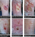
 At this point, a calcium alginate dressing impregnated with ALH was applied to both wounds. The dressings were changed twice weekly; within 1 week, Mr. J reported a decrease in pain to 4 out of 10 and his clinician noted the well-demarcated wound margin had diminished and become nearly flush with the surrounding periwound skin (see Figure 1 and Figure 2). Within 3 weeks, wound volume improved 73%. In 7 weeks, Mr. J’s pain level decreased to a 0 out of 10 and he reported an improved ambulation ability (see Figure 3). After 2.5 months, the wound had improved in volume (length x width x depth) by 56% (see Figure 4) and by 81% in 12 weeks (see Figure 5), with complete resolution in 16 weeks (see Figure 6).
As with most current wound care dressings, honey dressings are simple to apply, generally painless, do not harm the wound bed and tissues, create a moist wound environment, and because they are antibacterial, promote healing and epithelialization.4 These properties make ALH a viable alternative to other antimicrobial dressings in patients who cannot tolerate silver-containing dressings. In this case study, the ALH dressing was found to be effective in reducing wound pain and promoting granulation and epithelialization, resulting in complete wound healing.
Making Progress With Stalled Wounds is made possible through the support of Derma Sciences, Inc., Princeton, NJ. The opinions and statements of the clinicians contained herein are specific to the respective authors and are not necessarily those of Derma Sciences, Inc., OWM, or HMP Communications.
This article was not subject to the Ostomy Wound Management peer-review process.


