Preserved Left Atrial Function Following Left Atrial Appendage Closure for Stroke Prevention
Abstract
Background. Patients with atrial fibrillation (AF) are at high risk of thromboembolism, with most thrombi forming in the left atrial (LA) appendage. LA appendage closure is an alternative therapy to oral anticoagulation for stroke prevention in AF patients with contraindication to oral anticoagulation. LA function is critical for cardiovascular function, and recent studies suggested a direct relationship between LA function and AF recurrence. Deformation imaging characterizes and quantifies myocardial function. Aim. This study aims to investigate the impact of LA appendage closure on LA function in patients with paroxysmal AF. Methods. We studied patients with paroxysmal AF who underwent LA appendage closure in a single-center, retrospective study. Twelve patients (CHA2DS2-VASc score, 4.12 ± 1.1; age, 75.9 ± 6.9 years; 7 men and 5 women) were eligible. Echocardiography-derived LA global longitudinal strain analysis, LA diameter, and LA volume index were determined before and after a 6-month follow-up. All patients were in sinus rhythm during echocardiography. The LA global longitudinal strain was unchanged after LA appendage closure (from -18.9 ± 2.8% to -19.6 ± 2.6%; P=.66). No changes were observed for LA size (from 49.1 ± 6.1 mm to 50.5 ± 5.2 mm; P=.45) or for LA volume index (from 51.6 ± 4.6 mL/m² to 52.1 ± 4.1 mL/m²; P=.49), corroborating unaltered LA function after LA appendage closure. Conclusion. LA function is crucial for cardiovascular function and recurrence of AF. Our study provides evidence that LA appendage closure preserves LA function, determined by strain imaging in patients with paroxysmal AF and sinus rhythm during echocardiography.
J INVASIVE CARDIOL 2021;33(1):E40-E44. doi:10.25270/jic/20.00257
Key words: atrial fibrillation, LA appendage closure, thrombotic stroke
Atrial fibrillation (AF) is the most common cardiac arrhythmia, and patients with AF are at high risk of thromboembolism, with most thrombi originating from the left atrial appendage (LAA).1 The LAA is the source of embolic thrombus formation in more than 90% of cardiac thromboembolisms.2 Stroke prevention mainly necessitates the use of oral vitamin-K antagonists (VKAs) or non-VKA antagonists (novel oral anticoagulants). LAA closure is an alternative therapy to oral anticoagulation for stroke prevention in patients with AF, especially when oral anticoagulation is contraindicated.3-6 LA function is a critical contributor for cardiovascular diseases and plays a role in cardiac performance by modulating left ventricular function. LA remodeling is prognostically relevant for different diseases, and recent meta-analyses suggest a direct relationship between LA function and AF recurrence.7,8 Strain echocardiography is a valuable tool for assessment of LA remodeling and mechanics, and is able to detect subclinical LA dysfunction.9 Furthermore, global longitudinal strain (GLS) is a sensitive method for determination of myocardial functional recovery.10,11 Based on the fact that the LAA closure device is large compared with other interventional implantable devices and is placed in the LA wall, it remains unknown whether LAA closure alters LA function. Furthermore, no data regarding LA alterations exist in patients with paroxysmal AF and sinus rhythm during echocardiography following LAA closure.
The question arises as to whether LAA closure impacts LA function with improved or even worse remodeling. The present study thus aims to evaluate LA function and potential remodeling after LAA closure.
Methods
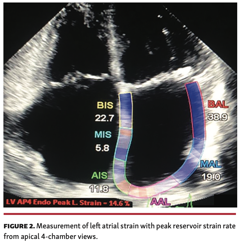
Design and study population. Between February 2015 and January 2018, a total of 302 patients were screened (Figure 1). Twelve patients with paroxysmal AF for >1 year with contraindications to oral anticoagulants and sinus rhythm during echocardiographic measurements and underwent LAA closure were enrolled in an open-label, single-center, observational, retrospective study. Before device implantation, LAA flow velocity and presence of LA thrombi were evaluated by transesophageal echocardiography (TEE). After femoral venous access, the LA was accessed by means of transseptal puncture in all patients. Experienced operators performed the LAA closures under TEE and fluoroscopy for guidance using dedicated devices (first-generation Amplatzer Cardiac Plug or second-generation Amplatzer Amulet; St. Jude Medical). Device oversizing of 3-5 mm was typically applied, as previously described.12 Patient and procedural characteristic imaging findings, laboratory results, in-hospital and follow-up data up to 6 months after LAA closure were collected in a dedicated database. TEE was performed before the procedure, periprocedurally, and during follow-up. Procedural success was defined as successful implantation of the device in the LAA without leak by postprocedure TEE. All patients were in sinus rhythm and underwent clinical assessment, conventional echocardiography, and GLS analysis of the LA at baseline and during a 6-month follow-up. The LA strain measurements were obtained from the two-dimensional (2D), apical, 4-chamber view and calculated with the negative peak strain rate during contraction phase and the reference lines for analysis were set on the P waves. The LA strain during contraction phase occurs during mitral valve closure in patients with sinus rhythm (Figure 2), as described previously.13
Study procedures were in accordance with the Declaration of Helsinki and the institutional ethics committee of the local university, which approved the study protocol (16-7080-BO). Patient records were deidentified and analyzed anonymously; therefore, the local ethics committee approved the retrospective analysis of the patient data without the need to obtain patient consent.
Statistical analysis. The data were expressed as mean ± standard deviation and compared using the paired t-test or Mann-Whitney U-test. Normal distribution was checked using the Kolmogorov-Smirnov test. Data analysis was performed with SPSS Statistics, version 22 (IBM for Windows). A P-value <.05 was considered statistically significant.
Results
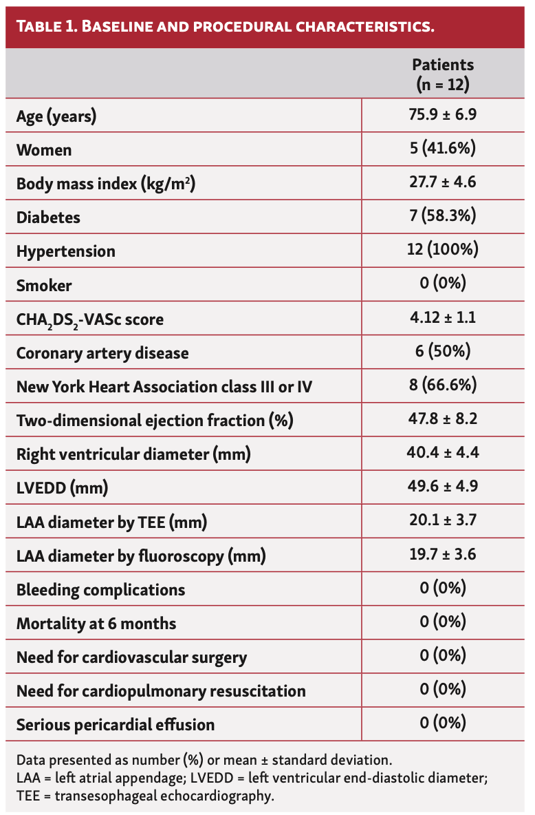
Table 1 lists the baseline characteristics of the 12 patients (age, 75.9 ± 6.9 years; 58.4% men) with indications for LAA closure enrolled with paroxysmal AF (CHA2DS2-VASc score, 4.12 ± 1.1) and sinus rhythm during echocardiography.
Overall procedural success and survival during follow-up were achieved in all patients with no complications. The device size used was between 16 mm and 30 mm (device size, 25.6 ± 4.9 mm). The overall procedure duration was 35.3 ± 9.21 minutes. No patients died or were lost to follow-up at 6 months, and no bleeding complications or stroke occurred. Oral anticoagulation was discontinued immediately following successful LAA closure and dual-antiplatelet therapy was initiated, consisting of 100 mg aspirin and 75 mg clopidogrel daily for 6 months, at the physician’s discretion. All patients took antihypertensive medication and were normotensive during measurements.
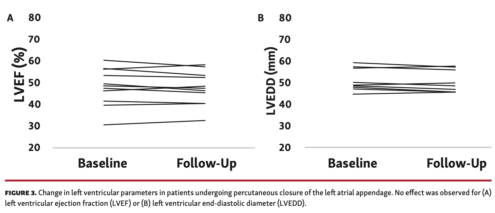
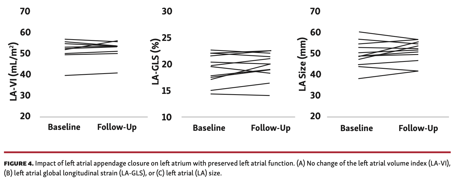
The goal of this study was to evaluate LA function and potential remodeling after LAA closure. To this aim, patients with paroxysmal AF and sinus rhythm during in-hospital presentation underwent clinical assessment, conventional echocardiography, and GLS analysis of the LA by speckle-tracking echocardiography at baseline and during a 6-month follow-up. Regarding left ventricular function, no change was observed after LAA closure for ejection fraction (from 47.8% ± 8.2% to 47.1% ± 7.4%; P=.66) (Figure 3A) or for left ventricular end-diastolic diameter (from 49.6 ± 4.9 mm to 48.2 ± 4.7 mm; P=.45) (Figure 3B). There was no difference after LAA closure in LA volume index (from 51.6 ± 4.6 mL/m² to 52.1 ± 4.1 mL/m²; P=.49) (Figure 4A), LA-GLS (-18.9 ± 2.8% to -19.6 ± 2.6%; P=.66) (Figure 4B), or LA size (49.1 ± 6.1 mm to 50.5 ± 5.2 mm; P=.45) (Figure 4C).
Discussion
Stroke is a significant cause of mortality in AF patients. The standard therapy to prevent stroke and peripheral embolization in AF is oral anticoagulation.1 Hemorrhagic complications preclude many patients from being prescribed or maintained on therapy, and the incidence of intracerebral hemorrhage in patients using oral anticoagulation will continue to increase with the demographic change of an aging population.14,15 As compared with primary spontaneous intracerebral hemorrhage, bleeding under oral anticoagulation is characterized by larger hematoma volumes with worse prognoses.16,17 Regarding thromboembolism, the appendage is the primary location of thrombi generation in 90% of LA clots.18
LAA closure is the valid alternative to anticoagulation in patients with AF and contraindications to anticoagulation for stroke prevention according to current guidelines.19,20 Recent trials showed even a beneficial effect of LAA closure in AF, with a similar stroke reduction and a significant reduction in major bleeding events including hemorrhagic stroke and consequently a lower mortality when compared with oral anticoagulation.21 AF is associated with LA enlargement and leads to remodeling of the LA. The LA dysfunction is associated with an increased risk for cardiovascular disease.22 The question arises as to whether LAA closure has an impact on LA function with potential improved or even worse remodeling. As shown in other trials, a potential impact of LAA closure on LA dimensions can be observed in patients with persistent AF.23
Our study is the first to investigate LA function secondary to LAA closure in patients with paroxysmal AF and sinus rhythm during echocardiography. This is significant, as preserved LA functions are increasingly important in heart failure, heart failure with preserved ejection fraction, and recurrence of AF.24-26 We have now identified unaltered dimensions and functions in LA volume index, LA size, and LA strain before and 6 months after LAA closure. LA strain is a useful parameter for evaluating LA function, is sensitive in estimating intracavitary pressures, and provides highly reproducible measures of LA deformation.27 There is a growing body of evidence supporting a strain echocardiography to use in different clinical settings.28-30 In our analysis, we found no change of LA-GLS, LA volume index, or LA size after LAA closure. Our findings therefore suggest that LAA closure potentially even preserves LA function in patients with paroxysmal AF >1 year and contraindications for oral anticoagulants. Interestingly, recent studies show that LAA closure is associated with an improvement in conduit, reservoir, and booster LA function, and no significant improvement was noted for strain indices for LA conduit function. Of note, a difference in our study is the Amplatzer devices implanted, which might potentially affect LA function. The Watchman occluder tends to stretch the LAA more than the Amplatzer Cardiac Plug or Amplatzer Amulet, which could lead to altered preload, causing an improvement in the conduit and reservoir function.31 Another study described improved LA mechanical function following LAA closure. However, LA strain parameters were calculated during the reservoir phase and measurements were conducted only 45 days after the procedure, while we present long-term data with a 6-month follow-up.32
Taken together, our results are important, as LAA closure has been shown to be non-inferior to oral antagonists for stroke prevention and has been proven even beneficial regarding bleeding complications. Of note, not only stroke prevention but also quality of life and cost effectiveness over a long-term follow-up are major determinants that play key roles when considering patients for LAA closure.33 Clearly, further larger-scale trials have to be conducted to observe a patient-relevant translation of our findings into clinical practice. The small sample size is the limiting factor in our study, mainly due to examinations during sinus rhythm in patients with paroxysmal AF.
Conclusion
LA function is crucial for cardiovascular functions in cardiovascular diseases, and recurrence of AF critically depends on LA performance. Our study provides the further evidence that interventional LAA closure preserves long-term LA function, determined by strain imaging in patients with paroxysmal AF and sinus rhythm during echocardiography.
From the Department of Cardiology and Vascular Medicine, West German Heart and Vascular Center Essen, University of Duisburg-Essen, Essen, Germany.
Disclosure: The authors have completed and returned the ICMJE Form for Disclosure of Potential Conflicts of Interest. The authors report no conflicts of interest regarding the content herein.
Final version accepted May 12, 2020.
Address for correspondence: Christos Rammos, MD, FESC, West German Heart and Vascular Center Essen, Department of Cardiology and Vascular Medicine, University Hospital Essen, Hufelandstraße 55, 45147 Essen, Germany. Email: christos.rammos@uk-essen.de
- Wolf PA, Abbott RD, Kannel WB. Atrial fibrillation as an independent risk factor for stroke: the Framingham study. Stroke. 1991;22:983-988.
- Qamruddin S, Shinbane J, Shriki J, Naqvi TZ. Left atrial appendage: structure, function, imaging modalities and therapeutic options. Expert Rev Cardiovasc Ther. 2010;8:65-75.
- Perales IJ, San Agustin K, DeAngelo J, Campbell AM. Rivaroxaban versus warfarin for stroke prevention and venous thromboembolism treatment in extreme obesity and high body weight. Ann Pharmacother. 2020;54:344-350.
- Kushnir M, Choi Y, Eisenberg R, et al. Efficacy and safety of direct oral factor Xa inhibitors compared with warfarin in patients with morbid obesity: a single-centre, retrospective analysis of chart data. Lancet Haematol. 2019;6:e359-e365.
- Yu LJ, Chen S, Xu Y, Zhang ZX. Clinical analysis of antithrombotic treatment and occurrence of stroke in elderly patients with nonvalvular persistent atrial fibrillation. Clin Cardiol. 2018;41:1353-1357.
- Lind A, Azizy O, Lortz J, et al. Percutaneous left atrial appendage closure for stroke prevention in patients with chronic renal disease. Archives of Medical Sciences. Epub Feb 25, 2020.
- Holmes DR Jr, Doshi SK, Kar S, et al. Left atrial appendage closure as an alternative to warfarin for stroke prevention in atrial fibrillation: a patient-level meta-analysis. J Am Coll Cardiol. 2015;65:2614-2623.
- Holmes DR Jr, Reddy VY. Left atrial appendage and closure: who, when, and how. Circ Cardiovasc Interv. 2016;9:e002942.
- Kowallick JT, Lotz J, Hasenfuss G, Schuster A. Left atrial physiology and pathophysiology: role of deformation imaging. World J Cardiol. 2015;7:299-305.
- Biering-Sorensen T, Hoffmann S, Mogelvang R, et al. Myocardial strain analysis by 2-dimensional speckle tracking echocardiography improves diagnostics of coronary artery stenosis in stable angina pectoris. Circ Cardiovasc Imaging. 2014;7:58-65.
- Karlsen S, Dahlslett T, Grenne B, et al. Global longitudinal strain is a more reproducible measure of left ventricular function than ejection fraction regardless of echocardiographic training. Cardiovasc Ultrasound. 2019;17:18.
- Azizy O, Rammos C, Lehmann N, et al. Percutaneous closure of the left atrial appendage in patients with diabetes mellitus. Diab Vasc Dis Res. 2017;14:407-414.
- Rammos C, Zeus T, Balzer J, et al. Left atrial and left ventricular function and remodeling following percutaneous mitral valve repair. J Heart Valve Dis. 2016;25:309-319.
- Sembill JA, Kuramatsu JB, Hohnloser SH, Huttner HB. Management of oral anticoagulation related intracerebral hemorrhage. Herz. 2019;44:315-323.
- Gokce E, Beyhan M, Acu B. Evaluation of oral anticoagulant-associated intracranial parenchymal hematomas using CT findings. Clin Neuroradiol. 2015;25:151-159.
- Biffi A, Kuramatsu JB, Leasure A, et al. Oral anticoagulation and functional outcome after intracerebral hemorrhage. Ann Neurol. 2017;82:755-765.
- de Schipper LJ, Baharoglu MI, Roos Y, de Beer F. Medical treatment for spontaneous anticoagulation-related intracerebral hemorrhage in the Netherlands. J Stroke Cerebrovasc Dis. 2017;26:1427-1432.
- Garcia-Fernandez MA, Torrecilla EG, San Roman D, et al. Left atrial appendage Doppler flow patterns: implications on thrombus formation. Am Heart J. 1992;124:955-961.
- Landmesser U, Holmes DR Jr. Left atrial appendage closure: a percutaneous transcatheter approach for stroke prevention in atrial fibrillation. Eur Heart J. 2012;33:698-704.
- Wintgens LIS, Vorselaars VMM, Klaver MN, et al. Left atrial appendage closure in atrial fibrillation patients with prior major bleeding or ineligible for oral anticoagulation. Neth Heart J. 2019;27:613-620.
- Kabra R, Girotra S, Vaughan Sarrazin M. Clinical outcomes of mortality, readmissions, and ischemic stroke among Medicare patients undergoing left atrial appendage closure via implanted device. JAMA Netw Open. 2019;2:e1914268.
- Lam CS, Rienstra M, Tay WT, et al. Atrial fibrillation in heart failure with preserved ejection fraction: association with exercise capacity, left ventricular filling pressures, natriuretic peptides, and left atrial volume. JACC Heart Fail. 2017;5:92-98.
- Phan QT, Shin SY, Cho IS, et al. Impact of left atrial appendage closure on cardiac functional and structural remodeling: a difference-in-difference analysis of propensity score matched samples. Cardiol J. 2019;26:519-528.
- Seewoster T, Kornej J. Left atrial function and NT-proANP as markers of AF progression and impaired outcome in patients with heart failure with preserved ejection fraction. Int J Cardiovasc Imaging. 2020;36:121-122.
- Morris DA, Ma XX, Belyavskiy E, et al. Left ventricular longitudinal systolic function analysed by 2D speckle-tracking echocardiography in heart failure with preserved ejection fraction: a meta-analysis. Open Heart. 2017;4:e000630.
- Reddy YNV, Obokata M, Egbe A, et al. Left atrial strain and compliance in the diagnostic evaluation of heart failure with preserved ejection fraction. Eur J Heart Fail. 2019;21:891-900.
- Morris DA, Takeuchi M, Krisper M, et al. Normal values and clinical relevance of left atrial myocardial function analysed by speckle-tracking echocardiography: multicentre study. Eur Heart J Cardiovasc Imaging. 2015;16:364-372.
- Collier P, Phelan D, Klein A. A test in context: myocardial strain measured by speckle-tracking echocardiography. J Am Coll Cardiol. 2017;69:1043-1056.
- Favot M, Courage C, Ehrman R, Khait L, Levy P. Strain echocardiography in acute cardiovascular diseases. West J Emerg Med. 2016;17:54-60.
- Ryczek R, Krzesinski P, Krzywicki P, Smurzynski P, Cwetsch A. Two-dimensional longitudinal strain for the assessment of the left ventricular systolic function as compared with conventional echocardiographic methods in patients with acute coronary syndromes. Kardiol Pol. 2011;69:357-362.
- Murtaza G, Vuddanda V, Akella K, et al. Impact of left atrial appendage occlusion on left atrial function — the LAFIT Watchman study. J Interv Card Electrophysiol. 2020;58:163-167.
- Coisne A, Pilato R, Brigadeau F, et al. Percutaneous left atrial appendage closure improves left atrial mechanical function through Frank-Starling mechanism. Heart Rhythm. 2017;14:710-716.
- Boersma LV, Ince H, Kische S, et al. Evaluating real-world clinical outcomes in atrial fibrillation patients receiving the WATCHMAN left atrial appendage closure technology. Circ Arrhythm Electrophysiol. 2019;12:e006841.


