ADVERTISEMENT
Left Atrial Appendage Occlusion in Patients With Thrombus in Left Atrial Appendage
Abstract: Background. Atrial appendage (LAA) occlusion is a therapeutic option for thromboembolic prevention in atrial fibrillation (AF) patients who have contraindications to oral anticoagulation (OAC) or high risk of bleeding. Traditionally, thrombus in the LAA has been considered a contraindication for LAA occlusion. Recently, resistant thrombus formation in patients using OACs was suggested as an indication for LAA occlusion. Methods and Results. In this single-center study, we evaluated the safety and efficacy of LAA occlusion in patients with a thrombus in the LAA. Twelve non-valvular AF patients who had a thrombus in the LAA were enrolled. The mean age was 71.8 years (range, 62-83 years). Permanent AF was present in all patients. Mean CHA2DS2-VASc score was 4.9 (range, 2-8) and mean HAS-BLED score was 4.8 (range, 3-6). Thrombi in the LAA were classified as type 1 (proximal to mid) and type 2 (distal) in 3 and 9 patients, respectively. Median follow-up duration was 12 months (interquartile range, 6-24 months). LAA occlusion was performed successfully with Amplatzer Amulet device without any significant periprocedural adverse events in all 12 patients. Transesophageal echocardiography (TEE) was performed at 1 and 6 months post procedure. Cardiovascular and all-cause mortality, significant ischemic cerebrovascular events, worsening heart failure, and major bleeding events did not occur during follow-up. Device-related thrombus was not observed with TEE in any patient. Conclusion. Our study showed that percutaneous LAA closure could be a therapeutic option for patients with resistant LAA thrombus.
J INVASIVE CARDIOL 2020;32(6):222-227. Epub 2020 April 24.
Key words: atrial fibrillation, LAA, LAA thrombus, left atrial appendage, percutaneous left atrial appendage closure
Atrial fibrillation (AF) is the most common sustained arrhythmia, with an increasing incidence with age.1 Ischemic stroke is a major problem in patients with AF. Oral anticoagulation (OAC) is the current gold standard therapeutic option for thromboembolic prevention in non-valvular AF patients.2 On the other hand, bleeding is an important complication of OAC treatment, and life-threating bleedings may occur in high-risk patients. The most common source of thrombus in patients with non-valvular AF is left atrial appendage (LAA).3 LAA occlusion is another therapeutic option for thromboembolic prevention in AF patients, especially in those with contraindications to OAC treatment or with high risk of bleeding.2
LAA occlusion is an intervention that is targeted to isolate LAA flow from the left atrium with a device.4 Clinical trials have demonstrated non-inferior outcomes for LAA occlusion compared with warfarin for thromboembolic prevention in non-valvular AF. 5 Bleeding complications are significantly reduced with LAA occlusion, because these patients are followed without OAC treatment. Classically, LAA thrombus is accepted as a contraindication to LAA occlusion.4 However, some case series showed that LAA occlusion could be performed safely and effectively in patients with thrombus formation in the LAA.6,7 Recently, resistant thrombus formation in patients using OAC was suggested as an indication for LAA occlusion in a consensus document.4 In this study, we therefore evaluated the safety and efficacy of LAA occlusion in patients with LAA thrombus.
Method
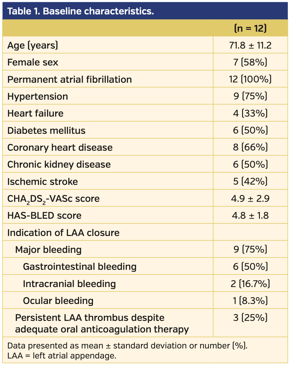
Study population. Eighty patients who had percutaneous LAA occlusion performed at Hacettepe University Cardiology Clinic between September 2015 and December 2018 were followed. Twelve of 80 non-valvular AF patients had thrombus in the LAA, and comprise the study population. Baseline characteristics, CHA2DS2-VASc score, HAS-BLED score, preprocedural and postprocedural antithrombotic medication, indications for LAA closure, and periprocedural adverse events were recorded. Patients were followed for 6 months for a significant clinical event, including stroke, cardiovascular mortality, and all-cause mortality. Transesophageal echocardiography (TEE) was performed at 1-month and 6-month follow-up for demonstration of any device-related thrombus and effective occlusion (peridevice leak <3 mm). Informed consent was obtained from all patients before enrollment. The study was approved by our institutional ethics committee (project number, GO 19/483 at the Hacettepe University local ethics committee).
Procedure. All patients were evaluated with transthoracic echocardiography (TTE) and TEE before the percutaneous LAA occlusion by the same cardiologist. LAA dimensions, thrombus dimensions, and location of the thrombus in the LAA cavity were evaluated.
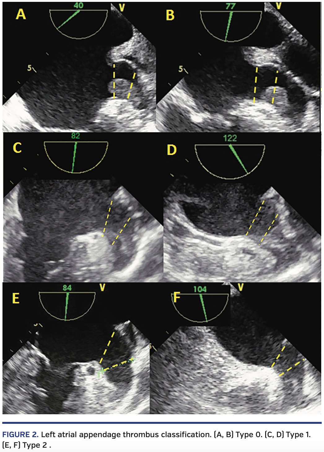
LAA thrombi were classified according to the location. The LAA is divided into three different sections (Figure 1). The first section is outside of the LAA. The second section is a 10 mm distance from the LAA ostium (landing zone). The third section is the base of the LAA. If any part of the thrombus is in the first section, it is classified as type 0 (overflowing thrombus). If any part of the thrombus is in the second section but not in the first section, it is classified as type 1 (proximal to mid thrombus). If thrombus is located in the third section and has no part in the second section, it is classified as type 2 (mid to distal thrombus) (Figure 2). Type 0 thrombi were excluded and LAA occlusion was not performed in these patients.
Patients with normal renal function underwent multislice cardiac computed tomography for optimal evaluation of LAA anatomy and size, as well as the relationship between the LAA and related cardiovascular structures.
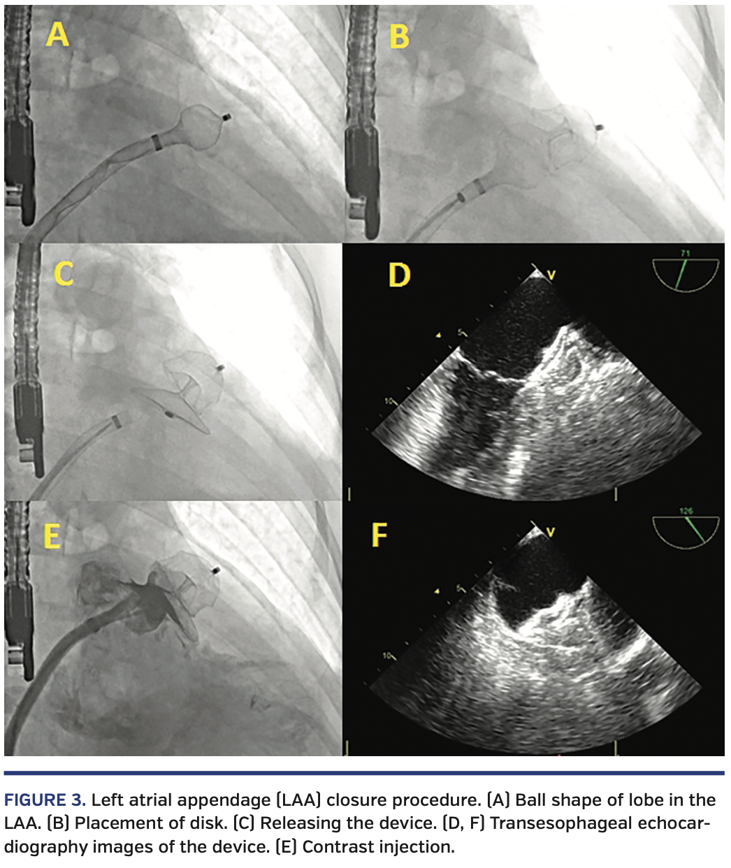
The percutaneous LAA occlusion procedure was performed under general anesthesia in all patients, and patients were intubated for optimal TEE guidance. Transseptal puncture was performed with fluoroscopy and three-dimensional (3D) TEE guidance. Transseptal puncture was performed at the inferoposterior site of the interatrial septum if there were no anatomic variations that prevented optimum orientation. After successful transseptal puncture, the delivery catheter was placed in the left atrium. An Amplatzer Amulet LAA occlusion device (Abbott Vascular) was advanced to some extent out of the delivery sheath in order to form a ball shape with the lobe of the device. The delivery sheath with the ball-shaped device on the tip was then placed in the LAA with counter-clockwise rotation, avoiding any unnecessary manipulation. The LAA was not engaged with a pigtail catheter and contrast injection was not performed to avoid embolization of the thrombus. All measurements and device size selections were based on TEE. After confirming the optimum settlement in the LAA with TEE, the lobe of the device was opened with further advancement. To prevent catheter-induced thrombus embolization, inappropriate catheter manipulation in the LAA was avoided. After proper placement of the lobe in the LAA, the disc was opened at the ostium of the LAA with withdrawal of the delivery sheath (Figure 3). The relationship between the occlusion device and the circumflex artery/mitral valve were checked with 3D-TEE and radiopaque contrast was injected within the delivery sheath to evaluate peridevice leak. Before releasing the device, it was pulled back with enough force to check the stability. The occlusion device was then released, and the relationships between the device and LAA, circumflex artery, and mitral valve were evaluated by 3D-TEE. Periprocedural anticoagulation was maintained by intravenous heparin infusion with activated clotting time monitoring.
Postprocedural antiplatelet therapy. The antiplatelet therapy post procedure was planned as either dual-antiplatelet therapy or a continuation of anticoagulant therapy. This individualized therapy was designed in accordance with the indication for LAA occlusion and each patient’s thromboembolism and bleeding risk.
Postprocedural follow-up. All 12 patients were re-evaluated clinically and with TTE at 1, 3, and the 6 months, while TEE was performed at 1 month and 6 months. Any adverse events, including bleeding complications, minor or disabling stroke, heart failure, and thromboembolic events, were recorded.
Statistical analysis. The statistical analysis was performed using SPSS statistical software, version 20 (SPSS). Descriptive and categorical variables were presented as frequencies and percentages. The continuous data with normal distribution are expressed as mean ± standard deviation. Quantitative variables without normal distribution are described with median and interquartile range. The condition of normality was evaluated using the Kolmogorov-Smirnov test. Numerical variables were compared using Student’s t-tests or Mann-Whitney U-test, as appropriate.
Results
Baseline characteristics. A total of 12 patients were enrolled. The mean age was 71.8 ± 11.2 years (range, 62-83 years). Permanent AF was present in all patients. Mean CHA2DS2-VASc score was 4.9 ± 2.9 (range, 2-8) and mean HAS-BLED score was 4.8 ± 1.8 (range, 3-6). Baseline characteristics are listed in Table 1. The main indication for LAA closure was a history of previous major bleeding (75%) while under OAC. Indication of the procedure in 3 patients (25%) was persistent LAA thrombus despite adequate OAC and antiplatelet therapy. Thrombi in the LAA were classified as type 1 and type 2 in 3 patients and 9 patients, respectively.
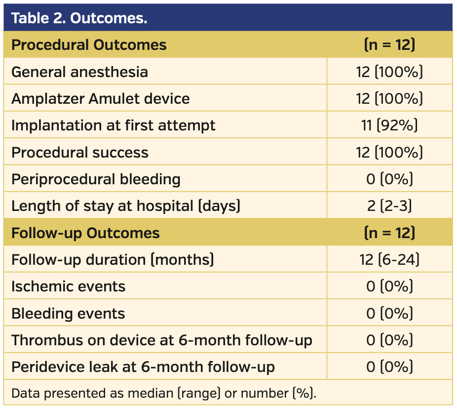
Procedural outcomes. Percutaneous LAA occlusion was performed successfully in all 12 patients. The Amplatzer Amulet device was used in all patients. The device was placed successfully during the first attempt in 11 of 12 patients. Cerebral protection device was not used in any patients. The mean in-hospital length of stay was 2.5 days (range, 2-4 days). Periprocedural major bleeding complications and significant ischemic cerebrovascular events were not observed in any patients. In 3 patients with persistent thrombus in LAA despite adequate anticoagulation, electrical cardioversion was performed and sinus rhythm was restored on day 1 post procedure. Procedural and follow-up outcomes were listed in Table 2.
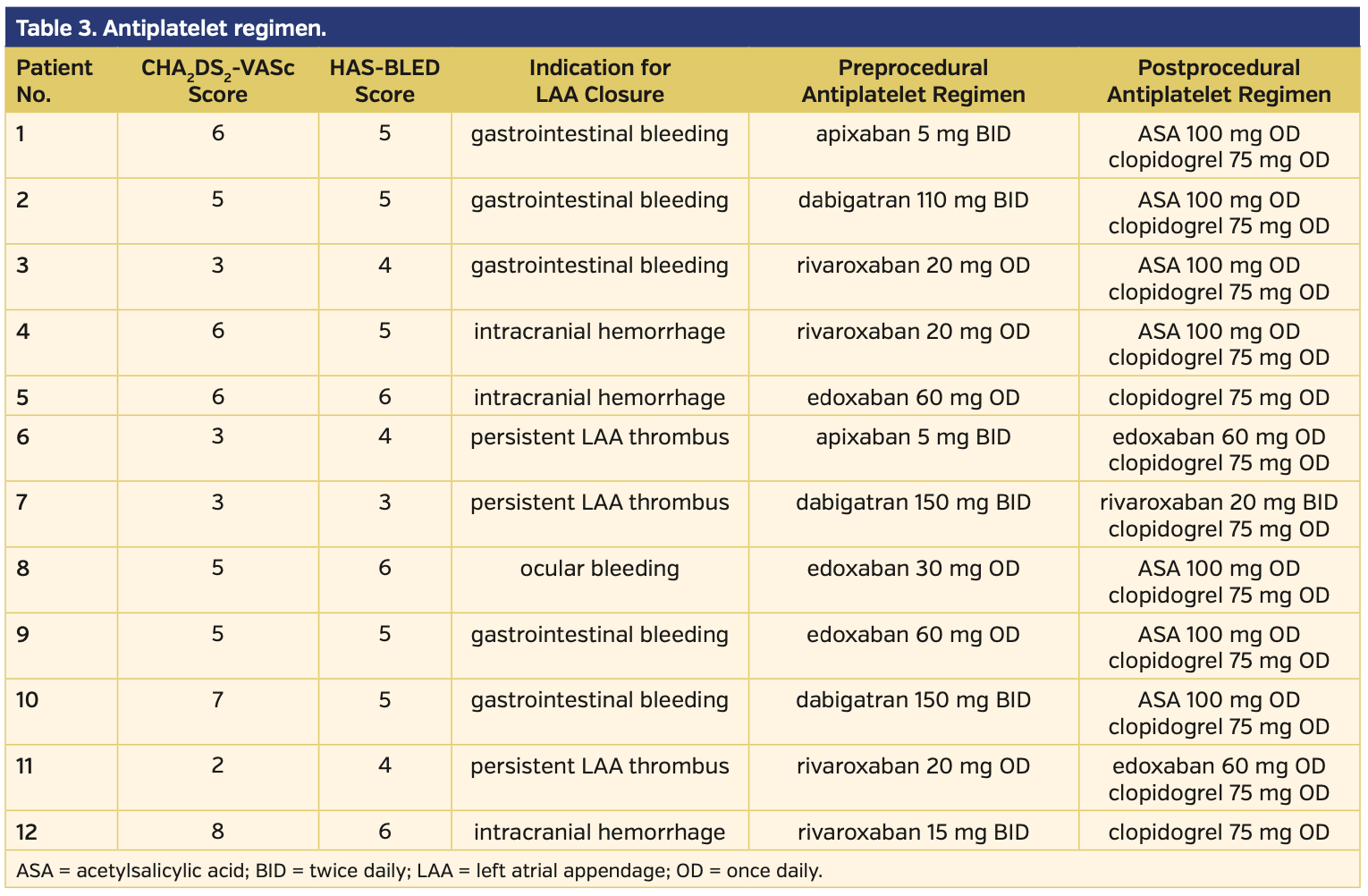
Follow-up. All patients were followed at 1 month, 3 months, and 6 months after the procedure. Median follow-up duration was 12 months (range, 6-24 months). Cardiovascular and all-cause mortality, significant ischemic cerebrovascular events, worsening heart failure, and major bleeding events were not observed during follow-up. Postprocedural antiplatelet/anticoagulant therapy was decided according to baseline characteristics and indications for the procedure. Preprocedural and postprocedural antithrombotic regimen and changes during follow-up are listed in Table 3.
TEE was performed 1 month and 6 months after the procedure in all patients. Device-related thrombus was not observed in any patients. There was no significant peridevice leak (>3 mm) in any of the 12 patients.
Discussion
Thrombus formation in the LAA was traditionally considered to be a contraindication to percutaneous LAA closure.8 Consequently, patients with thrombus in the LAA were excluded from large-scale percutaneous LAA closure studies.9 However, as the experience of the operators improved over time, case reports about LAA occlusion in patients with a thrombus in the LAA were published by various authors. We previously reported the first case of our series of LAA occlusion in patients with LAA thrombus.6 To the best of our knowledge, it was the first case with the Amulet device. Recently, Tarantini et al reported results of LAA occlusion in 32 patients with LAA thrombus in their multicenter series.10 In this series, percutaneous LAA closure was performed successfully in all patients and significant ischemic events were not reported during follow-up. There was only 1 device-related thrombus in the 23 patients who underwent TEE. Similarly, our study showed excellent technical and procedural success rates for LAA closure in patients with LAA thrombus. In our series, periprocedural major bleeding or ischemic complications were not observed. Postprocedural ischemic events were not reported during follow-up and no device thrombus was observed over 6 months of TEE follow-up.
In our study, the Amplatzer Amulet device was used in all cases. The Amulet is a self-expandable nitinol device with two parts (a lobe and a disc) premounted on a single cable.11 The external disc provides more appropriate coverage of the LAA ostium. This may create an advantage to avoid embolization during procedure. There are cases of LAA closure with other devices in the literature. Randomized trials are needed to compare the safety and efficacy of devices in patients with LAA thrombus.
In our series, we refined the Amplatzer Amulet implantation technique to avoid embolization of the thrombus in the LAA during the procedure. First, we took maximum precautions to avoid any engagement of wires or catheters in the LAA. Contrast injection with pigtail catheter was not performed and measurements were based on TEE. The device delivery catheter with the ball-shaped device on its tip was advanced into the LAA with a single maneuver in a “one shot” manner. After demonstrating the appropriate position in the LAA with TEE, the device lobe was opened with further advancement, without any pullback in the delivery catheter. Finally, all 80 procedures, including the 12 with LAA thrombus, were performed by the same operators and cardiovascular imaging specialist in this single-center series. We believe that this is a major factor for the excellent procedural outcomes reported in this study.
Antiplatelet regimens of patients were individualized according to patient characteristics. To date, there has been no consensus about long-term antiplatelet regimen after LAA occlusion.8 As we know, a major thrombus source in non-valvular AF is LAA.3 Consequently, if the patient had no contraindications to OAC and the indication for LAA closure was persistent LAA thrombus, we continued OAC after the procedure. In our series, patients used clopidogrel alone or dual-antiplatelet therapy with acetylsalicylic acid and clopidogrel, or continued using OAC in addition to single antiplatelet agent (acetylsalicylic acid or clopidogrel). This individualized antithrombotic regimen may have an impact on reducing thrombus formation on the device. We observed no device-related thrombus on TEE at follow-up.
Persistent thrombus in the LAA despite adequate OAC is an important concern in patients with AF. The thrombus in LAA precludes cardioversion in these patients, and this may be an especially significant problem in patients having rapid ventricular response. Embolic stroke despite adequate OAC and presence of thrombus in the LAA is another major condition that signifies the need for an alternative therapeutic option.4 In our series, we performed LAA occlusion in 3 patients with persistent thrombus in the LAA despite adequate anticoagulation. The procedure was successfully performed in these patients and sinus rhythm was restored with electrical cardioversion. AF ablation with pulmonary vein isolation was performed in these patients in a different session. In a recent consensus document, thromboembolic event or documented presence of thrombus in the LAA despite adequate OAC therapy was considered to be a potential indication for LAA occlusion.4
Cerebral protection device was not used in any patients in our series because of reimbursement issues. However, an interventional radiologist experienced in stroke intervention is ready on-site in our catheterization laboratory in case a periprocedural embolic cerebrovascular event occurs. We did not observe any ischemic cerebrovascular events during the procedure or at follow-up. It is possible that clinically non-significant cerebral or systemic embolization could have occurred. However, we did not perform any cerebral imaging to demonstrate microemboli. Clinically non-significant emboli can be observed with imaging modalities in AF patients despite adequate anticoagulant therapy.12
Our study has some strengths. First, it was designed as a single-center prospective study. All procedures were performed by the same operators who are experienced in LAA occlusion as well as other structural heart interventions. As such, operator-related complication diversity was minimal, compared with multicenter studies. Patients were closely followed, and TEE was performed in all patients by the same experienced cardiologist.
Study limitations. This was a non-randomized, observational study. Follow-up duration was relatively short. Although we did not observe any procedural complications or ischemic embolization during follow-up, the study was not designed and adequately powered to show clinical efficacy and safety of LAA occlusion in patients with LAA thrombus compared with OAC therapy. To demonstrate clinical success of LAA occlusion in this specific patient group, large-scale randomized studies are needed.
Conclusion
This study showed that percutaneous LAA closure in patients with a thrombus in LAA can be performed safely and efficiently by experienced operators. Percutaneous LAA closure may be an option for patients who have thrombus in the LAA despite adequate OAC therapy. Ischemic cerebrovascular events were not reported during follow-up. To verify the efficacy and safety of LAA closure in this patient group, large-scale randomized studies are needed.
From the Hacettepe University, Ankara, Turkey, and Ankara City Hospital Department of Cardiology, Ankara, Turkey.
Disclosure: The authors have completed and returned the ICMJE Form for Disclosure of Potential Conflicts of Interest. The authors report no conflicts of interest regarding the content herein.
The authors report that patient consent was provided for publication of the images used herein.
Manuscript submitted October 3, 2019, final version accepted October 18, 2019.
Address for correspondence: Cem Coteli, MD, Ankara City Hospital, Cardiology, Üniversiteler Mahallesi Bilkent Cad. No:1 Çankaya/ANKARA, Ankara, Çankaya 06800, Turkey. Email: cemcoteli@hacettepe.edu.tr
- Chugh SS, Havmoeller R, Narayanan K, et al. Worldwide epidemiology of atrial fibrillation: a Global Burden of Disease 2010 study. Circulation. 2014;129:837-847.
- Kirchhof P, Benussi S, Kotecha D, et al. 2016 ESC guidelines for the management of atrial fibrillation developed in collaboration with EACTS. Eur Heart J. 2016;37:2893-2962.
- Bajwa RJ, Kovell L, Resar JR, et al. Left atrial appendage occlusion for stroke prevention in patients with atrial fibrillation. Clin Cardiol. 2017;40:825-831.
- Tzikas A, Holmes DR Jr, Gafoor S, et al. Percutaneous left atrial appendage occlusion: the Munich consensus document on definitions, endpoints, and data collection requirements for clinical studies. Europace. 2017;19:4-15.
- Bajwa RJ, Kovell L, Resar JR, et al. Left atrial appendage occlusion for stroke prevention in patients with atrial fibrillation. Clin Cardiol. 2017;40:825-831.
- Aytemir K, Aminian A, Asil S, et al. First case of percutaneous left atrial appendage closure by amulet™ device in a patient with left atrial appendage thrombus. Int J Cardiol. 2016;223:28-30.
- Cammalleri V, Ussia GP, Muscoli S, De Vico P, Romeo F. Transcatheter occlusion of left atrial appendage with persistent thrombus using a trans-radial embolic protection device. J Cardiovasc Med (Hagerstown). 2016;17:e224.
- Meier B, Blaauw Y, Khattab AA, et al. EHRA/EAPCI expert consensus statement on catheter-based left atrial appendage occlusion. Europace. 2014;16:1397-1416.
- Fountain RB, Holmes DR, Chandrasekaran K et al. The PROTECT AF (WATCHMAN Left Atrial Appendage System for Embolic PROTECTion in Patients with Atrial Fibrillation) trial. Am Heart J. 2006;151:956-961.
- Tarantini G, D’Amico G, Latib A, et al. Percutaneous left atrial appendage occlusion in patients with atrial fibrillation and left appendage thrombus: feasibility, safety and clinical efficacy. EuroIntervention. 2018;13:1595-1602.
- Tzikas A, Gafoor S, Meerkin D, et al. Left atrial appendage occlusion with the AMPLATZER Amulet device: an expert consensus step-by-step approach. EuroIntervention. 2016;11:1512-1521.
- Berman JP, Norby FL, Mosley T, et al. Atrial fibrillation and brain magnetic resonance imaging abnormalities. Stroke. 2019;50:783-788.


