Hypertrophic Obstructive Cardiomyopathy Requiring Ablation via Septals From Both Left and Right Coronary Arteries — A Case Report and Review of Literature
Abstract: Alcohol septal ablation has traditionally been performed using septal perforators from the left coronary system. We describe a case in which septal perforators from the left and right coronary arteries were utilized and review current literature on the management of hypertrophic obstructive cardiomyopathy.
Key words: alcohol septal ablation, hypertrophic obstructive cardiomyopathy
Case Presentation
A 60-year-old woman with hypertrophic obstructive cardiomyopathy (HOCM) was referred for alcohol septal ablation (ASA) due to exertional dyspnea despite optimal medical management. Echocardiography showed a mean left ventricle-aorta pressure gradient of 105 mm Hg and severe mitral regurgitation due to systolic anterior motion of the mitral valve (SAM).
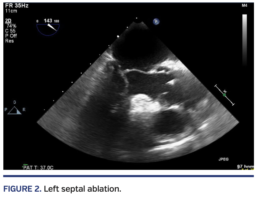
Left heart catheterization confirmed the presence of left ventricle-aorta pressure gradient (Figure 1) and showed septal perforators originating from the proximal segment of the right coronary artery (RCA), as well as the left anterior descending artery (LAD) (Videos 1 and 2). A guiding catheter was used to engage the left main coronary artery, and a coronary wire was passed into the LAD septal perforator. An over-the-wire balloon was then advanced. Contrast injection into the balloon catheter showed enhancement of the subaortic part of the septum on transesophageal echocardiography (TEE) (Figure 2). Alcohol was then injected. Postablation Doppler echocardiography showed a residual left ventricle-aorta pressure gradient of 40 mm Hg with no improvement in SAM. The guiding catheter was then withdrawn.
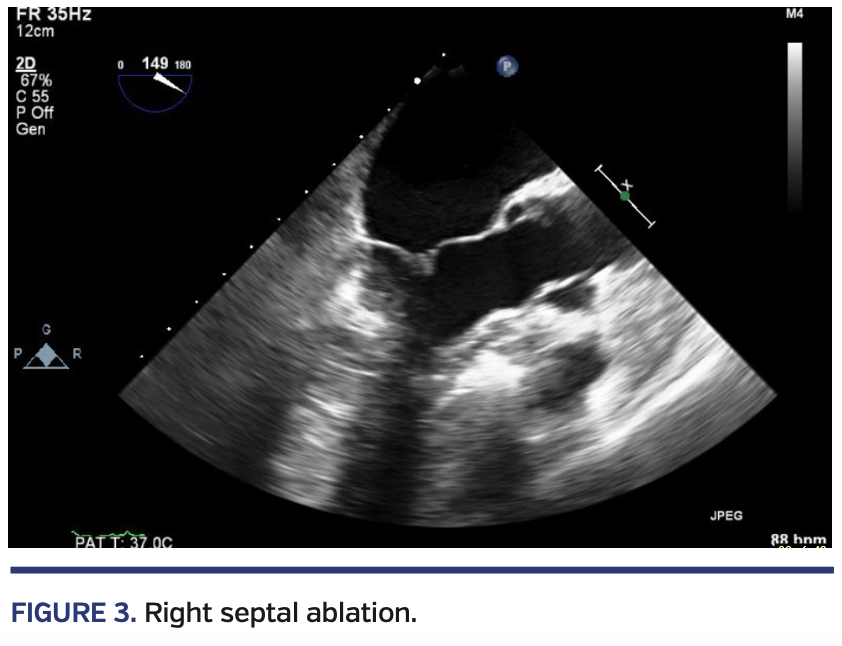
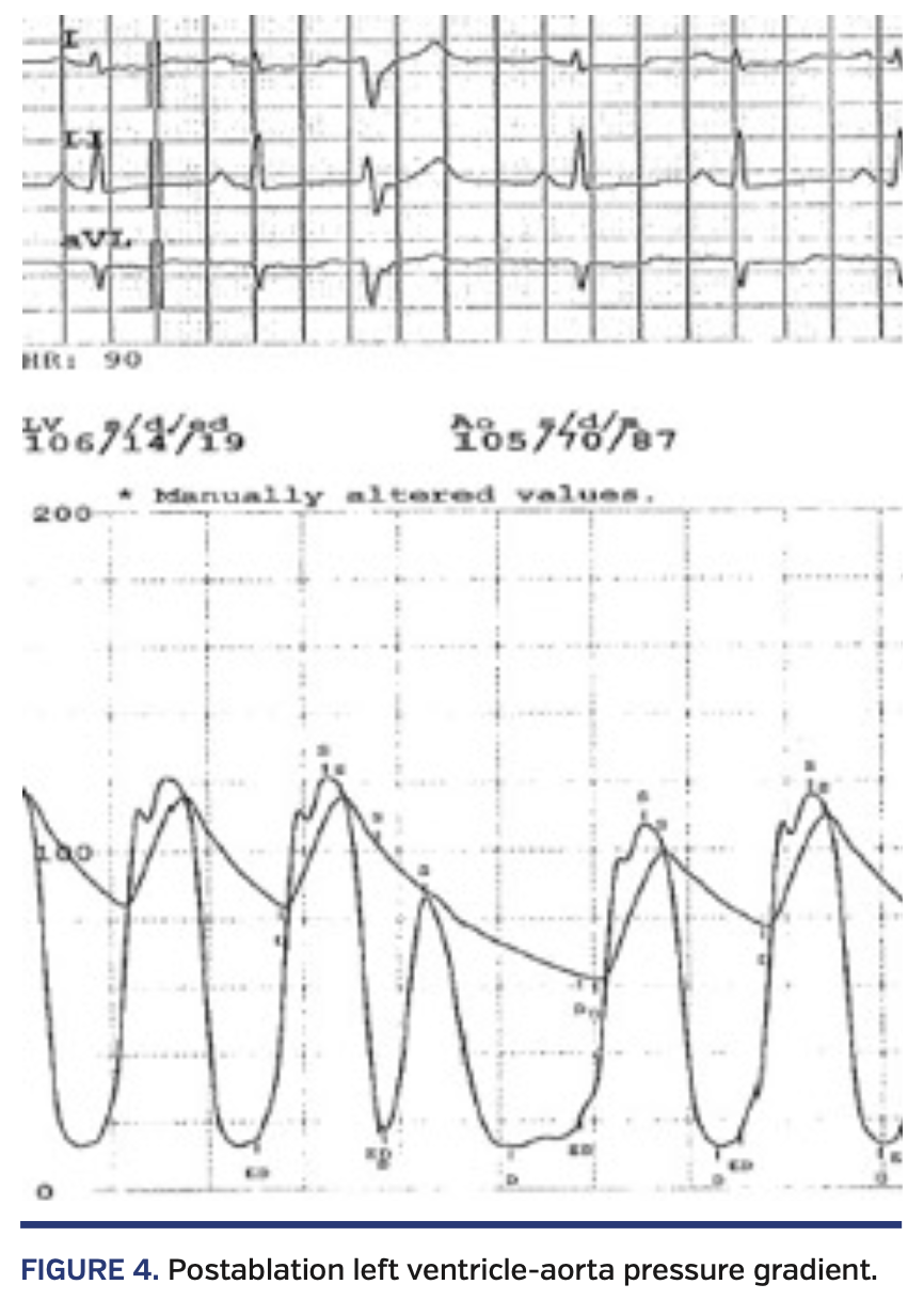
Via an RCA guiding catheter, a coronary wire was then advanced into the RCA and an over-the-wire balloon was advanced into the septal perforator. Contrast injection showed echocardiographic enhancement of a more apical segment of the septum (Figure 3). Additional alcohol was administered after the balloon was inflated. Final Doppler echocardiography showed reduction of left ventricle-aorta pressure gradient to 5 mm Hg, which was consistent with catheter gradient (Figure 4). Mitral regurgitation appeared mild and SAM was resolved.
Discussion
HOCM is a genetic disorder characterized by left ventricular hypertrophy that is unexplained by secondary causes of hypertrophy, and a nondilated left ventricle with preserved or increased ejection fraction.1 It is the most common heritable cardiomyopathy, affecting 1 of 500 people.2
The hallmark of HOCM is the presence of left ventricular hypertrophy (LVH) with accompanying myofibrillar disarray. Myocardial fibrosis results in some degree of diastolic dysfunction in virtually all patients with HOCM.3
Dynamic left ventricular outflow tract (LVOT) obstruction, defined as LVOT gradient ≥30 mm Hg, is present in 70% of HOCM patients, and can occur at rest or with provocation.4 Septal-preponderant hypertrophy resulting in abnormal subvalvular mitral apparatus can lead to turbulent flow that drags the redundant mitral valve into the LVOT, resulting in decreased forward flow and SAM-mediated mitral regurgitation.5 The LVOT obstruction is associated with development of symptoms and increased cardiac morbidity and mortality.6
Clinical presentation of HOCM can range widely. The most common symptoms are shortness of breath and chest pain. Sudden cardiac death (SCD) is present in 0.5%-2% per year.1 Risk factors for SCD include unexplained syncope, hypotensive response to exercise, interventricular septum or ventricular wall thickness of ≥30 mm, a family history of HOCM with SCD, multiple episodes of documented non-sustained ventricular tachycardia, and extensive (≥15% of the left ventricular mass) late gadolinium enhancement determined by cardiac magnetic resonance imaging.1 Implanted cardioverter/defibrillator (ICD) is recommended for primary prevention in patients with one or more risk factors, and for secondary prevention in survivors of cardiac arrest thought to be due to HOCM.7
Pharmacologic therapy is directed at symptomatic relief, largely by reducing LVOT obstruction.5 The mainstay of therapy is beta-blockers, which have a negative inotropic effect therefore reducing dynamic LVOT obstruction, whereas its negative chronotropic effect prolongs diastole and allows for more left ventricular filling.7 Non-dihydropyridine calcium-channel blockers can be used when beta-blocker use is contraindicated. Disopyramide, a class Ia antiarrhythmic with negative inotropic effects, can be used alone or in conjunction with other therapies in patients who remain symptomatic despite adequate atrioventricular blockade, and can be used with a beta-blocker or a non-dihydropyridine calcium-channel blocker in patients with LVOT obstruction and atrial fibrillation, as long as left ventricular systolic function is preserved.7,8
Septal reduction therapy is reserved for patients with refractory symptoms despite optimal medical management. Surgical myectomy is the gold standard of septal reduction according to American College of Cardiology Foundation/American Heart Association guidelines7 and is favored in younger patients with less comorbidities. ASA carries a class IIa recommendation in patients with LVOT obstruction and severe drug-refractory symptoms when surgery is contraindicated or the risk is considered unacceptable because of serious comorbidities or advanced age,7 or as an alternative to surgical myomectomy in eligible patients (class IIb recommendation). The European Society of Cardiology guidelines provide comparable recommendations to ASA, unless patients have other indications for open-heart surgery, in which case surgical myomectomy is preferred.9
The operative mortality of surgical myomectomy is reported at 0.3%-1.1%.10 In experienced centers, intermediate-term mortality following ASA is comparable to that after surgical myectomy.11,12 ASA frequently results in right bundle-branch block, whereas myectomy creates a left bundle-branch block.5 Pacemaker dependency post ASA occurs in 10%-15% and is most likely to occur in patients with pre-existing left bundle-branch block.13 There are no randomized controlled trials comparing surgical myomectomy to ASA.5
Conclusion
If present, septal perforators from the right coronary artery can be safely used for ASA when desired results cannot be achieved by utilizing the left coronary artery septal perforators.
From the Department of Cardiology, Newark Beth Israel Medical Center, Newark, New Jersey.
Disclosure: The authors have completed and returned the ICMJE Form for Disclosure of Potential Conflicts of Interest. The authors report no conflicts of interest regarding the content herein.
Manuscript accepted February 12, 2020.
Address for correspondence: Mohammed Alomar, MD, 12902 Magnolia Drive, MCB-CPT, Tampa, FL 33612-9416. Email: mohammedalomar@yahoo.com
- Marian AJ, Braunwald E. Hypertrophic cardiomyopathy: genetics, pathogenesis, clinical manifestations, diagnosis, and therapy. Circ Res. 2017;121:749-770.
- Semsarian C, Ingles J, Maron MS, Maron BJ. New perspectives on the prevalence of hypertrophic cardiomyopathy. J Am Coll Cardiol. 2015;65:1249-1254.
- Ommen SR, Nishimura RA. Hypertrophic cardiomyopathy. Curr Probl Cardiol. 2004;29:239-291.
- Maron MS, Olivotto I, Zenovich AG, et al. Hypertrophic cardiomyopathy is predominantly a disease of left ventricular outflow tract obstruction. Circulation. 2006;114:2232-2239.
- Geske JB, Ommen SR, Gersh BJ. Hypertrophic cardiomyopathy: clinical update. JACC Heart Fail. 2018;6:364-375.
- Maron MS, Olivotto I, Betocchi S, et al. Effect of left ventricular outflow tract obstruction on clinical outcome in hypertrophic cardiomyopathy. N Engl J Med. 2003;348:295-303.
- Gersh BJ, Maron BJ, Bonow RO, et al. 2011 ACCF/AHA guideline for the diagnosis and treatment of hypertrophic cardiomyopathy: a report of the American College of Cardiology Foundation/American Heart Association Task Force on Practice Guidelines. Developed in collaboration with the American Association for Thoracic Surgery, American Society of Echocardiography, American Society of Nuclear Cardiology, Heart Failure Society of America, Heart Rhythm Society, Society for Cardiovascular Angiography and Interventions, and Society of Thoracic Surgeons. J Am Coll Cardiol. 2011;58:e212-e260.
- Sherrid MV, Barac I, McKenna WJ, et al. Multicenter study of the efficacy and safety of disopyramide in obstructive hypertrophic cardiomyopathy. J Am Coll Cardiol. 2005;45:1251-1258.
- Elliott PM, Anastasakis A, Borger MA, et al. 2014 ESC guidelines on diagnosis and management of hypertrophic cardiomyopathy: the task force for the diagnosis and management of hypertrophic cardiomyopathy of the European Society of Cardiology (ESC). Eur Heart J. 2014;35:2733-2779.
- Maron BJ, Dearani JA, Ommen SR, et al. Low operative mortality achieved with surgical septal myectomy: role of dedicated hypertrophic cardiomyopathy centers in the management of dynamic subaortic obstruction. J Am Coll Cardiol. 2015;66:1307-1308.
- Agarwal S, Tuzcu EM, Desai MY, et al. Updated meta-analysis of septal alcohol ablation versus myectomy for hypertrophic cardiomyopathy. J Am Coll Cardiol. 2010;55:823-834.
- Sorajja P, Ommen SR, Holmes DR Jr, et al. Survival after alcohol septal ablation for obstructive hypertrophic cardiomyopathy. Circulation. 2012;126:2374-2380.
- Maron BJ, Nishimura RA. Surgical septal myectomy versus alcohol septal ablation: assessing the status of the controversy in 2014. Circulation. 2014;130:1617-1624.


