Vascular Ring and Recurrent Pneumonia
An 11-year old girl presented with history of recurrent pneumonia since early childhood and symptoms suggestive of gastroesophageal reflux. She was sent for cardiac evaluation as a part of a workup for persistent respiratory symptoms.
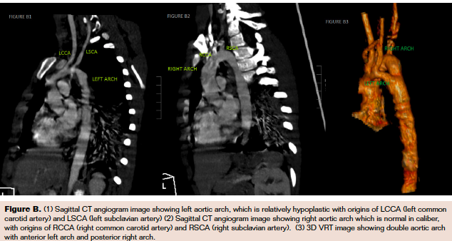
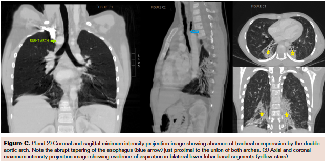
 Her physical examination and electrocardiogram results were essentially unremarkable. Chest x-ray was suggestive of right aortic arch with consolidation in the right lower zone. Echocardiogram did not reveal any intracardiac lesion but raised suspicion of abnormality of aortic arch—presence of 4 neck vessels instead of the typical 3 vessels. Computed tomography (CT) angiogram was subsequently done, and it revealed the presence of double aortic arch (Figure A), with a larger right-sided arch (blue arrow) and smaller left arch (green arrow) joining to form the descending aorta (grey arrow). The right subclavian and common carotid arteries arose from the right arch (Figure B2) and, similarly, left subclavian and common carotid arteries from the left arch (Figure B1). A sudden taper (blue arrow in Figure C2) in the esophageal caliber was noted, suggesting esophageal compression, which may have exacerbated the respiratory symptoms.
CT also showed bilateral lower lobe consolidation (yellow stars in Figure C3). In view of her symptoms, surgery is planned with an aim to resect the lesser of the arches. Vascular rings should be considered in patients with persistent respiratory symptoms in the absence of any intracardiac lesion, after an initial workup for recurrent pneumonia.
Disclosure: The authors have completed and returned the ICMJE Form for Disclosure of Potential Conflicts of Interest. The authors report no conflicts of interest regarding the content herein.
Manuscript submitted October 10, 2017; accepted October 16, 2017.
Address for correspondence: Priyadarshini Arunakumar, DM; Sree Chitra Tirunal Institute for Medical Sciences and Technology, Trivandrum, India. Email:. priya_arun_2000@yahoo.com


