Advanced Signal Acquisition and Analysis Help Drive Procedural Efficiency and Efficacy
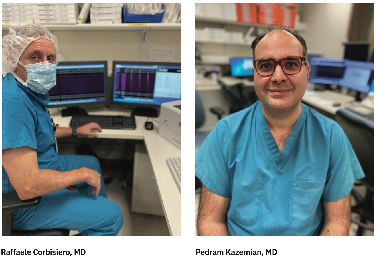
In this feature interview, EP Lab Digest speaks with Raffaele Corbisiero, MD and Pedram Kazemian, MD about their use of the PURE EP™ System (BioSig Technologies, Inc.) at the Deborah Heart and Lung Center in Browns Mills, New Jersey, as they are currently evaluating the technology.
Q&A with Raffaele Corbisiero, MD
Can you give us your impression about the integration of the PURE EP™ System with the other equipment you have in your EP lab?
The integration is seamless, so it doesn’t affect anything that we have set up already or anything else that we do. So that’s nice.
Having a seamless integration allowed us to really focus on the most important thing, which is the physiologic information that we get from the signals. When we started using the PURE EP™ System, we had a setup giving us signals from the PURE EP™ system and from our recording system, we immediately noticed that we really got more signal information from PURE EP™ than we did from our conventional systems.
As we get down the road, and as our experience will increase, I can see how precise signals will become very important for our practice, across all ablation procedures.
What are the key shortcomings that the PURE EP™ system is addressing?
It’s really about the amount of information that we get from the electrical signals, because a lot of times, especially in our labs, we get a lot of noise. The PURE EP™ System has a low-noise design and provides complete flexibility, allowing me to determine what is real, and what is not real. So that kind of information is really important and gives me more confidence during my ablations.
Can you describe the impact PURE EP™ has on your clinical decision-making process?
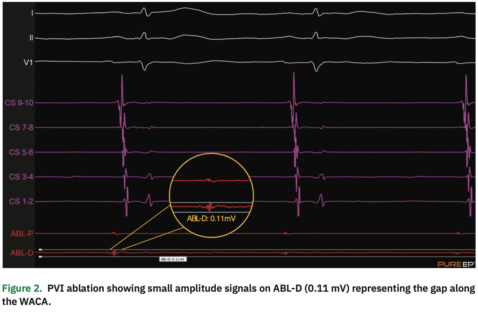
As far as my clinical decision-making process, I’ve noticed since adding the PURE EP™ System that during PVIs, I have a better sense of when I’ve completely isolated the veins. I’ve observed a higher occurrence of “first-pass” pulmonary vein isolations since using the PURE EP™ system. When the PV potentials on PURE EP™ go flat, I feel more confident I’m done with that ablation, so it has actually increased my ablation efficiency.
How do you see the value of PURE EP™ in contributing to the overall procedural efficiency/efficacy?
Having more accurate signals will translate into more effective and efficient ablation procedures. That is really the key. As we go forward with more complex ablations, especially redos and VTs, I think the signal quality is going to matter immensely. You have to be able to see what you’re doing, and to me, the PURE EP™ System has opened my eyes to signals I couldn’t appreciate before.
Q&A with Pedram Kazemian, MD
Can you tell us more about the quality of the signals you can obtain with PURE EP™ and how the additional information adds value to your clinical practice?
Because of the superior quality of the signals, the one distinctive feature of it which is different from our current recording system, is the significantly less background noise that we often see in our labs.
I can see 3 general areas where PURE EP™ can be incorporated into my practice.
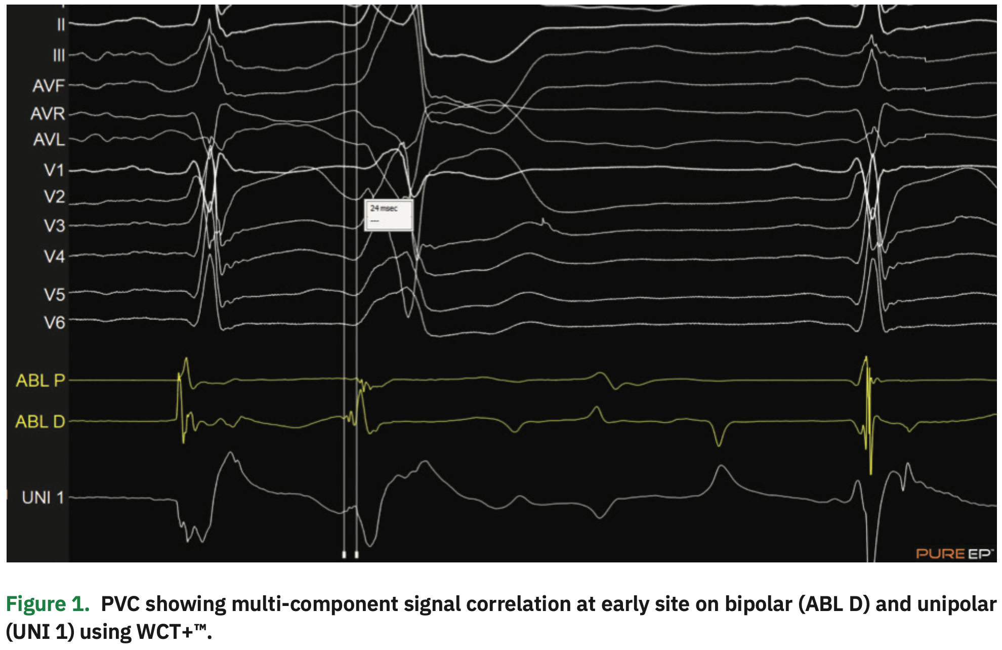
The first one is in terms of mapping and localization of tachycardias, especially within the scar. Most atrial and ventricular tachycardias are secondary to scar reentry, resulting in low-voltage local EGMs that are often buried within the far field signals of the surrounding tissue. This creates a challenge for activation mapping. Anything that improves the signal-to-noise ratio helps us create more accurate maps. We can manually adjust and annotate the local activation in the electroanatomic map based on the measurement of the local activation in the PURE EP™ compared to a reference EGM.
A potential future application of PURE EP™ could be around finding endpoints for ablation. The question that we always face is whether we have ablated enough at any particular location to achieve transmural ablation. Since we can acquire very clean and stable unipolar signals using WCT+™, the reduction of signal amplitude or the conversion to a QS morphology could represent a possible endpoint for ablation. So for example, in AF ablation, we can continue radiofrequency delivery until we get reduction of signal by 70-80%. We have to develop those endpoints, so there is some research needed to find the appropriate physiologic information for ablation in the atrium and ventricle. But this opens the door to very exciting potential applications.
The third area where PURE EP™ may be useful would be arrhythmia diagnosis. For example, in AVNRT and AVRT, sometimes we are looking for a slow pathway potential or accessory pathway potentials that are not easy to detect. Furthermore, during pacing maneuvers, important diagnostic signals may be buried inside the saturation artifact from the pacing electrode. The wide dynamic range of the PURE EP™ system may allow us to better differentiate those signals as there is no system saturation and a quicker recovery to baseline.
How do you see the value of specialty digital filters such as the ‘high-frequency filter’ in contributing to the overall procedural efficiency/efficacy?
It is my understanding these filters work like an advanced equalizer within the PURE EP™ System, so we can eliminate undesired frequencies, especially if we already know the type of signals we’re looking for. One obvious clinical application is easier visualization of the His signal — this might particularly be useful in the field of His pacing.
Another application is when we have a lot of fractionation, especially in the scar of the atrium. Again, some of these fractionations blend into the background noise because we usually have to gain up on conventional systems to see lower voltage electrocardiograms. When we do this, the signal-to-noise ratio goes so low, everything becomes fuzzy and blends together. This is not the case with the PURE EP™ System, which needs no gain and is capable of differentiating signal components within complex electrograms. PURE EP™ can differentiate the high frequency component, which may be far-field. This capability may assist us with localizing the activation more precisely.
Can you describe a moment during a recent procedure where you felt that the signals you observed on PURE EP™ were unique and influenced the course of the procedure itself?
Yes. Oftentimes, during PVI procedures, it can be difficult to find areas of connection or reconnection, because we just ablated around the pulmonary veins where the signals are very low voltage. Using the PURE EP™ System during PVI procedures, I was able to achieve “first-pass” isolation. PURE EP™ was very helpful in identifying those areas.
There was also another case of a patient with an LVAD. Typically with LVADs, because of the electric motor, you get a lot of high-frequency noise on the 12-lead electrocardiogram. PURE EP™ was used to clear up the 12-lead electrocardiogram and eliminate the LVAD noise, while preserving other important physiologic signal information.
This article is published with support from BioSig Technologies, Inc.
Disclosures: The authors have no conflicts of interest to report regarding the content herein.


